Playlist
Show Playlist
Hide Playlist
Layers of the Heart Wall
-
Slides 01 Human Organ Systems Meyer.pdf
-
Reference List Histology.pdf
-
Download Lecture Overview
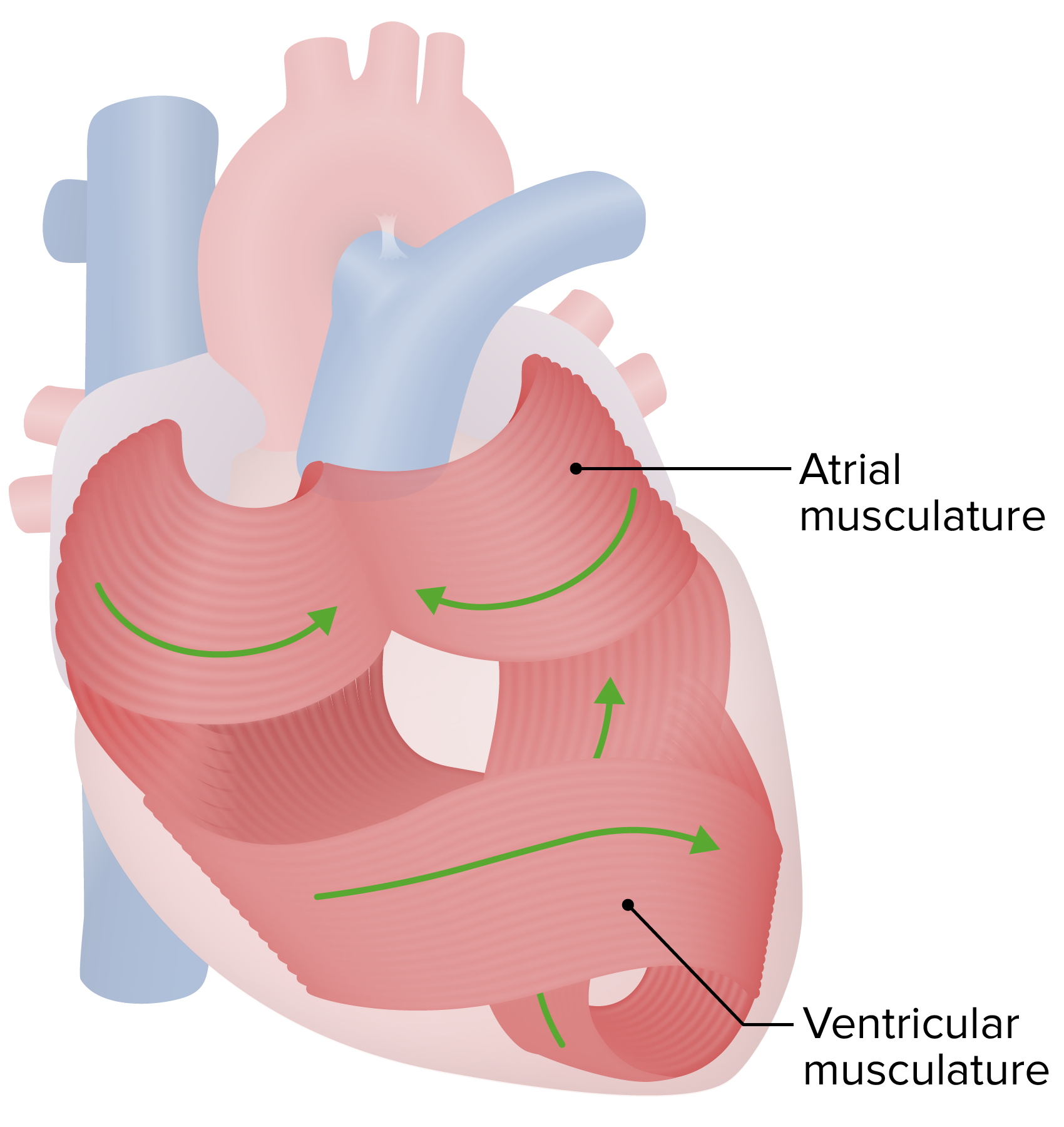
00:01 So, that's the sequence of blood flow through the heart. 00:04 Remind yourself of the physiology of the heart and the pumping of the heart, the atria contracting together, and the ventricles contracting together. 00:15 Now, let's now look, again, at this diagram and understand that there are three different components to the wall of the heart. 00:28 First of all, the heart is lined on the outside by a thin capsule or thin membrane called the epicardium. 00:38 This epicardium is actually a visceral layer of the pericardium. 00:44 Because that epicardium reflects back on itself and creates a pericardial cavity or sac in which the heart sits and in which the heart pumps. 00:55 And that cavity, that serous cavity is lined by fluid. 01:00 It's a very thin cavity, it's lined by fluid that reduces the friction during the beating of the heart. 01:07 So, the epicardium's on the outside. The thickest layer is the myocardium. 01:14 The myocardium is really full of cardiac muscle. That's the work part of the heart wall. 01:22 That's the part of the heart wall that does the contracting or the pumping, the myocardium. 01:29 And then, internally, the endocardium lines the entire inner surface of the heart. 01:36 It lines the heart valves as well and the septum between the atria and also, both ventricles called the interatrial and interventricular septums. 01:50 So, now, let's look at those structures in a bit more detail. 01:57 Let's look at the histology of those structures. 02:00 First of all, the epicardium, the covering on the outside of the heart. 02:05 As you can see from the diagram and also, from the histological section, it contains a lot of fatty tissue or adipose tissue and I'm sure those of you who have been in a dissection lab always find that rather frustrating when you're trying to locate some of the positions of the coronary arteries. 02:28 It's also composed of some connective tissue, collagen, elastic tissue, all the sorts of normal connective tissue fibers you'd expect to find in some loose connective tissue. 02:41 And on the outside, it's lined by a squamous type epithelium called the mesothelium. 02:49 So, that mesothelium, underlying connective tissue, and adipose tissue create the outside coating or covering of the heart, the epicardium. 03:00 The myocardium as I mentioned before, consists entirely of cardiac muscle with some connective tissue skeletal components, and also, blood vessels. 03:13 But it's mostly cardiac muscle and as I've said before, that's the pump component of the heart. 03:21 It's lined on top, of course, by endocardium. It's lined on the top of the section here but remember, it's lining the internal chambers of the heart. 03:33 So, have a look at the epithelium of the heart lining here, endothelium, and it consists of a squamous epithelial cell lining as I was mentioned before and that squamous epithelium sits on a subendothelial layer of connective tissue. 03:54 It's a very thin layer of supporting connective tissue, similar to a lamina propria in other organ systems. 04:01 And sometimes, there's also a third layer, the subendocardial layer, again, of connective tissue and this separates the epithelial surface from the underlying cardiac muscle. 04:15 And in some way, protects the delicate endothelial surface of the heart from the rather harsh, vigorous activity of the pumping cardiac muscle. 04:26 Well, one of the things you can't really appreciate when you look at histological sections of the heart, is that it actually does have a very strong fibrous skeleton and that fibrous skeleton, very dense connective tissue, dense collagen, helps to, well, it doesn't help but is the insertion point for all the cardiac muscle, so it's a very important component. 04:53 On this slide, you could see a tiny little section taken through the myocardium of the atrium and also, you can see that there's a septum of connective tissue between both atria here and also, between the ventricles. 05:10 Here's a section here showing you a label of a piece of myocardium of the ventricle. 05:18 Notice though that in between this myocardium of the atrium and myocardium of the ventricle, there is this clear component. 05:27 That clear component is part of the connective tissue, fibrous skeleton of the heart. 05:34 It's a membrane that separates the atria from the ventricle, and that's very important because that then stops the wave of impulse, and therefore, the wave of contraction going directly from the atrium muscle to the ventricle muscle. 05:52 So, it allows a delay, and, of course, that delay is very important between the filling of the ventricle and the contraction of the atrium. 06:03 So, it's really an electrical isolation sort of tissue there. It's very important.
About the Lecture
The lecture Layers of the Heart Wall by Geoffrey Meyer, PhD is from the course Cardiovascular Histology.
Included Quiz Questions
The visceral layer of the serous pericardium is best regarded as which of the following?
- The epicardium
- The endocardium
- The fibrous pericardium
- The myocardium
- The layer that maintains the integrity of the heart valves
Which of the following layers is the thickest layer of the heart?
- Myocardium
- Epicardium
- Endocardium
- Interatrial septum
- Serous pericardium
In the adult heart, fully differentiated white adipose tissue can be commonly found in which layer?
- Subepicardium
- Myocardium
- Interatrial septum
- Interventricular septum
- Endocardium
Where is the fibrous skeleton of the heart located?
- Between the atria and ventricles
- Between the atria
- Between the ventricles
- Around the atria
- Around the ventricles
Customer reviews
5,0 of 5 stars
| 5 Stars |
|
1 |
| 4 Stars |
|
0 |
| 3 Stars |
|
0 |
| 2 Stars |
|
0 |
| 1 Star |
|
0 |
I loved his lecture . He makes it very simple to understand and he focus on the important things .


