Playlist
Show Playlist
Hide Playlist
Larynx

-
Slides Anatomy Larynx.pdf
-
Reference List Anatomy.pdf
-
Download Lecture Overview
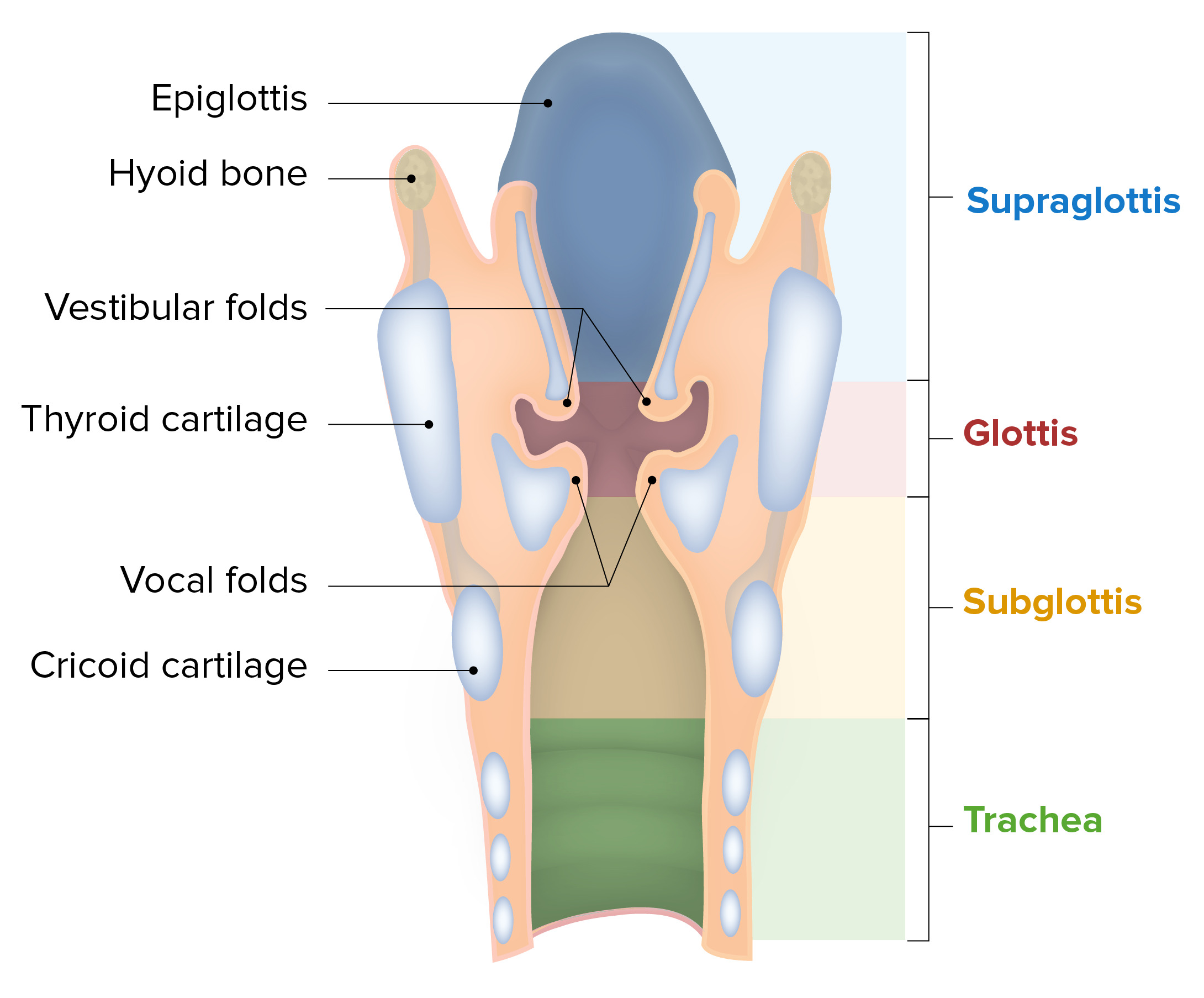
00:01 Let's finish off the neck by looking at the larynx. 00:05 We'll start with a very clinical view. 00:08 So this would be the type of view you have of the opening into the larynx, if you're doing a laryngeal scope. 00:17 Anteriorly, we would have the tongue Just behind that would be the epiglottis, a very important flap of tissue. 00:27 And there'd be a space between the tongue and the epiglottis. 00:30 That's called the molecula. 00:34 Looking down into the larynx itself, we would see two folds. 00:38 One that's more pink and lateral, and that will be the vestibular fold, also called the false vocal cords, And then projecting more medially, sitting below the vestibular folds would be the vocal folds, also called the true vocal cord. 00:53 And it has this more whitish appearance because of its participation in vocalization, and the need for a different type of mucosa. 01:05 The space between the vocal cords is called the glottis. 01:08 In this case, there is an open glottis because we can see through the vocal cords. 01:14 Posteriorly, we have another set of depressions. 01:19 Here we call them the piriform recess back in the pharyngeal area. 01:26 Here's a sagittal cross section to show the various levels of the larynx. 01:32 Superiorly, we have the supraglottic space. 01:35 In the area of the glottis we have the transglottic space, and then inferiorly the subglottic space before we enter the trachea. 01:45 Now let's look at what we call the laryngeal skeleton, which is composed of several cartilages. 01:51 Although with age, a lot of these cartilages will actually ossify in form partially at least bone. 01:58 Inferior to the larynx we have the cartilages of the trachea. 02:03 Just before we reach the trachea are inferior most cartilage of the larynx is the cricoid cartilage. 02:11 The largest is something called the thyroid cartilage Superiorly we have attachments to the hyoid bone Connecting the thyroid cartilage and the hyoid bone is this tough membrane called the thyrohyoid membrane. 02:28 But it has a very important opening or foramen that allows a branch of the superior laryngeal artery and nerve to go into that branch that pierces in is the internal branch. 02:41 We also have a membrane connecting the thyroid and cricoid cartilage is called the cricothyroid membrane. 02:47 And this is a soft area within the larynx that can be targeted during a cricothyroidotomy. 02:55 Also in this area connecting the two is the cricothyroid muscle, which is a very important muscle in the process of vocalization. 03:04 Now, let's swing around for a posterior view of the laryngeal skeleton. 03:09 Here we can see the thyroid cartilage but we see that the thyroid cartilage doesn't go all the way around. 03:15 It really is just an anterior structure shaped somewhat like a shield, which is actually what thyroid means. Means shield like. 03:23 The cricoid, on the other hand, is circumferential. 03:26 And does go all the way around the airspace. 03:30 We also see the cartilage of the epiglottis here. 03:33 As well as these pyramidal shaped cartilages called the arytenoids that sit on top of the cricoid cartilage. 03:41 There are somewhat extended by these smaller cartilages the corniculate and cuneiform cartilages Here we see the cricoid cartilage. 03:51 Again, going all the way around the airway. Unlike the thyroid. 03:56 Anteriorly, we have a small arch and then posteriorly we have these wider walls called the lamina. 04:03 At the top, we have these little facets for articulation with those arytenoid cartilages and they're the ones that are going to move during the process of vocalization. 04:14 We also facet for articulation with the inferior horn of the thyroid cartilage. 04:20 Here is that thyroid cartilage where it has some walls called the right and left lamina. 04:26 And where they meet inferiorly we have a notch called the inferior thyroid knotch. 04:31 Similar one superiorly called the superior thyroid notch. 04:35 And just below this, at the anterior most projection is something called the laryngeal prominence. 04:41 Something that's often palpable during physical exam and sometimes also called an Adam's apple. 04:48 Or posteriorly, we have a superior and inferior tubercle connected by an oblique line to which many structures will attach. 04:57 We also have that inferior horn for an articulation with the cricoid cartilage, and a superior horn to which ligaments will attach. 05:07 Here's a superior view of the thyroid cartilage, where we can again see the inferior and superior horns. 05:14 And we see that the lamina form an angle to call the thyroid angle. 05:18 And it varies between 90 to 120 degrees. 05:23 And actually the narrower angle creates more of an elongation and that is actually what will have an effect on the overall depth or pitch of a voice. 05:36 Here we see those pyramidal shaped arytenoid cartilage sitting on top of the cricoid. 05:42 They have a broad base inferiorly and a pointy aspect superiorly called the apex. 05:50 Anteriorly, we have a projection called the vocal process because it will interact with the vocal cords, then posteriorly a muscular process because that's where we're going to have attachments for these laryngeal muscles. 06:03 Here's another superior view of the laryngeal skeleton showing our thyroid cartilage anteriorly and the cricoid posteriorly. 06:12 They're connected at least anteriorly by this cricothyroid membrane. 06:17 We have the arytenoid cartilages sitting on top of the cricoid cartilage. 06:23 And here we find this ligament running from the arytenoids to the thyroid cartilage called the vocal ligaments. 06:30 Superior to which are a different set of ligaments called the vestibular ligaments. 06:38 Here we have another cross section view where we see the epiglottis, the hyoid, the thyroid cartilage anteriorly, the cricoid cartilage circumferentially, and the membrane between the two the cricothyroid membrane. 06:54 Again, the arytenoid sit superior to the cricoid cartilage. 07:00 The superior fold is the false vocal cord why the inferior one is the true vocal cord. 07:07 The one that actually produces vocalization. 07:11 Let's look at the muscles that produce that vocalization. 07:15 First of all, we have the posterior cricoarytenoid muscle, which is very descriptive in where it is. 07:22 We have lateral cricoarytenoid, transverse arytenoids, oblique arytenoids. 07:30 The vocalis running more anterior to posterior, as well as the thyroid retinoids which are also running more or less in that same direction. 07:40 Further laterally, we have muscle called the aryepiglotticus. 07:45 We also have the thyroepiglotticus. 07:49 When it comes to the movement, vocal cords can be abducted or brought away from the midline or adducted. 08:01 Here we see them abducting or opening versus adducting or closing. 08:10 When it comes to the cricothyroid muscle, it's actually drawing down that thyroid cartilage and an anterior inferior direction which is actually tensing those vocal cords creating a higher pitch. 08:28 Here we see the posterior cricoarytenoids which are very important because they're really the only muscles that cause abduction of the vocal cords. 08:39 And that's important not so much in terms of vocalization, but in terms of opening up the airway, because closed vocal cords would block air from getting down into the trachea and lungs. 08:52 We also have the lateral cricoarytenoids which are going to cause abduction. 09:01 the transverse arytenoids which are also going to cause abduction. 09:06 Then we have the vocalis and thyroid arytenoid which are oriented very differently. 09:12 They're more of an anterior posterior orientation along the length of the vocal cords. 09:18 And so they're going to adjust the tension in these vocal folds. 09:25 Now let's look at the blood supply of the larynx. 09:30 Here we see the superior laryngeal artery and vein and the inferior laryngeal artery, as well as the inferior laryngeal vein. 09:39 These are all branches off of the corresponding thyroid arteries and veins. 09:45 Innervation of the larynx really comes from cranial nerve X, or the vagus nerve. 09:50 And there is a bit of asymmetry between left and right. 09:52 So we'll start by looking at the left vagus nerve. 09:55 The vagus nerve has a branch called the superior laryngeal nerve, which will form an internal branch that will become internal by piercing through the thyrohyoid membrane. 10:07 And then we'll have an external branch. 10:09 And that external branch is going to innervate the cricothyroid muscle. 10:15 Here, we also see the relationship of the common carotid artery as it comes off of the arch of the aorta directly. 10:23 And that's a bit of a symmetry because the arch of the aorta is something of a leftward structure. 10:29 And because it's on the left side, the recurrent laryngeal nerve has to arch under the arch of the aorta much further down than it does on the right side. 10:41 Once it does, it will come back upwards and provide the motor innervation for the remainder of the laryngeal muscles. 10:51 On the right, we see, again the right vagus nerve. 10:55 But on the right side, the right recurrent laryngeal nerve will recur underneath the right subclavian artery. 11:03 So it will make that turn much more superiorly than it does on the left side of the body.
About the Lecture
The lecture Larynx by Darren Salmi, MD, MS is from the course Upper Aerodigestive Tract.
Included Quiz Questions
What lies between the tongue and the epiglottis?
- Vallecula
- Vestibular fold
- Vocal fold
- Piriform recess
- Glottis
Where is the foramen for the superior laryngeal artery located?
- Thyrohyoid membrane
- Hyoid bone
- Thyroid cartilage
- Cricoid cartilage
- Trachea
What is another name for Adam's apple?
- Laryngeal prominence
- Left lamina
- Right lamina
- Inferior thyroid notch
- Superior thyroid notch
What is a typical thyroid angle?
- 100 degrees
- 40 degrees
- 150 degrees
- 210 degrees
- 300 degrees
What runs between the thyroid cartilage and the arytenoid cartilage?
- Vocal ligament
- Cricoid cartilage
- Cricothyroid membrane
- Annular process
- Hyoid bone
Which of the following is NOT a laryngeal muscle?
- External arytenoid
- Transverse arytenoid
- Lateral cricoarytenoid
- Posterior cricoarytenoid
- Oblique arytenoid
Which movements are possible with the vocal cords? Select all that apply.
- Adduction
- Abduction
- Internal rotation
- External rotation
- Extension
Author of lecture Larynx

Darren Salmi, MD, MS
Customer reviews
5,0 of 5 stars
| 5 Stars |
|
2 |
| 4 Stars |
|
0 |
| 3 Stars |
|
0 |
| 2 Stars |
|
0 |
| 1 Star |
|
0 |
Really informative and good explanation with really nice illustrations min 10 words required
Very illustrative animation, the lecturer is also very clear and concise!
