Playlist
Show Playlist
Hide Playlist
Introduction to Spinal Cord

-
Slides Diseases of the Spinal Cord.pdf
-
Download Lecture Overview
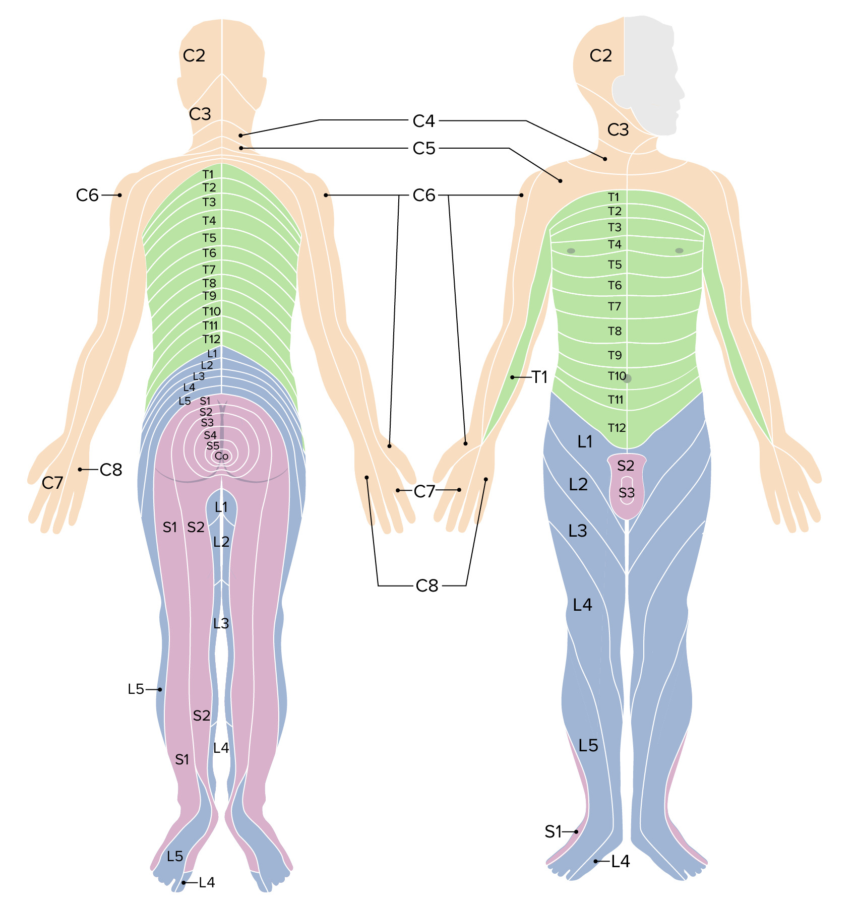
00:00 So let's talk about some introductory and orientation to the spinal cord. When we think about the spinal cord, we divide it into 4 segments. There is the cervical, thoracic, lumbar, and sacral segments and you can see those here. Each of these segments contains both gray matter and white matter tracts. The gray matter contains the cell bodies of the motor and then the key input tracts for the sensory fibers at each level of the spinal cord, and that here is depicted in pink. The yellow are the white matter tracts that surround the gray matter in the spinal cord. And there are a few things to take away from this schematic. First of all, there's a different amount of gray matter in each area and segment of the spinal cord. We have a lot of cell bodies in the cervical region and so more gray matter and more cell bodies arising out of that area to give our arms all of the innervation that they need. There is less gray matter in the thoracic spine where there are only a small number of nerve roots going to the thoracic body in that area. Down in the lumbar spine which gives rise to all the nerves leaving the lumbar spine to the legs and arriving in from the legs, we again see more gray matter. 01:12 And so you can see the variation in the amount of gray to white matter in each area of the spinal cord. Here, we're looking at the spinal cord laterally and there are a few things to take away from this. The vertebral bodies of the spine extend all the way from the top of the head or the base of the head and the skull all the way down to the end of the buttocks, but the spinal cord typically ends higher than that. We see the cervical, thoracic, lumbar, and sacral segments with vertebral bodies anterior to the spinal cord and the spinous processes posterior to the spinal cord. And importantly, we see the end of the spinal cord typically in adults around the L1 to L2 area which is called the conus medullaris, that end of the spinal cord, and from there the spinal cord gives rise to the cauda equina where those horsetail of peripheral nerves that descend in the vertebral bodies and exit out into the legs. And we can see pathology affecting the spinal cord proper from the upper cervical region C1 down to the upper lumbar region L1-2, and then we can see central nervous system pathology give rise to peripheral nervous system disorders by affecting the cauda equina. And those would be lesions in the lower lumbar space L2, 3, 4, and then down in the sacrum. So we're dealing with the spinal cord, we're dealing with the central nervous system, but within the vertebral bodies and the vertebral apparatus we have both central and peripheral nodes. Let's look at one segment, a representative segment of the spinal cord. 02:47 When we think about the spinal cord, we divide it into each individual segment and here you can see one schematically depicted. In each segment of the spinal cord, we have a ventral portion and a dorsal portion. The ventral portion of the spinal cord gives rise to the ventral nerve roots and there we see the fibers for the motor nerves that descend out from that segment of the spinal cord. And correspondingly, the dorsal nerve roots carry sensory information in from that segment of the body, that dermatome of the body. Those cells bodies arise and leave in the dorsal root ganglion and then travel in to the spinal cord where they either synapse or cross over to the opposite side of the spinal cord before ascending to the brainstem. We can also think about and look at the spinal cord topographically and here we're looking at a typical segment from top down and there are a number of important layers and areas of the spinal cord that will present differently. First, we see the spinal nerves inside the canal, the spinal cord proper. Just outside of the spinal cord is the subarachnoid space and that's very important. We need to see that space on MRI scans to know that there is non-compression or pathology that is affecting the spinal cord. Within the subarachnoid space, we see the nerve roots, the dorsal and ventral nerve roots as they exit or enter the spinal cord. Just lateral to those are the dorsal root ganglion which actually lie within the neural foramen, the foramen through which the nerves exit and are susceptible and sensitive to compression in that area. Then we have the dura which surrounds the dorsal root ganglia and then following that and outside of that is the epidural space and we can see pathology affecting the epidural space and compressing inwards to the spinal cord resulting in pathology. So when we think about the spinal cord, we can divide it into each of those areas and that can help us to understand typical clinical presentations for patients. The first location or place within the spinal cord we think about is the spinal cord proper, the nerves themselves. There is gray matter nuclei and white matter tracts. And certain pathology will affect the gray matter nuclei, things like strokes in the spinal cord or toxic metabolic problems within the spinal cord. There is also certain pathology that will affect the white matter tracts. In certain degenerative conditions as well as compressive lesions and various inflammatory or infectious processes can affect the white matter or gray matter. The second area within the spinal cord is the arteriovenous system. And we have arteries and in particular the anterior spinal artery will be very important for patient presentations from vascular problems affecting the spinal cord and the epidural venous plexus, that plexus around the spinal cord and the epidural space. And metastasis or infections, osteomyelitis, and others can affect the epidural space and present with a problem compressing the cord externally. The third area or region within the spinal cord are the vertebral bodies. Around the vertebral bodies we have anterior and posterior ligaments which can be susceptible to trauma or can affect the location of herniated disc or other degenerative pathology. We can see thickening of those ligaments compressing the spinal cord and causing myelopathic presentations. There is also the intervertebral disc material and in patients with degenerative disease or degeneration arthritis of the spinal cord, that disc can protrude and result in compression of the spinal cord contributing to patient's symptoms. And then the last area of the spinal cord and spinal region are the peripheral nerves, and that's that cauda equina, peripheral nerve that live inside the central nervous system and can be susceptible to pathology that that can present rather suddenly and should be on a clinician's differential diagnosis for some in many of these patients.
About the Lecture
The lecture Introduction to Spinal Cord by Roy Strowd, MD is from the course Diseases of the Spinal Cord.
Included Quiz Questions
What is the approximate level where the spinal cord terminates in most adults?
- Between L1 and L2
- Between L3 and L4
- Between L4 and L5
- Between L2 and L3
- Between T12 and L1
What term is used to describe the tapered lower end of the cord?
- Conus medullaris
- Cauda equina
- Coccygeal ligament
- Pia mater
- Filum terminale
In the spinal cord, the fibers of the motor neurons exit via the...
- ...ventral nerve roots of the spinal nerves.
- ...dorsal roots of the spinal nerves.
- ...ventral nerve roots in the cervical segment and the dorsal roots in the lumbar segment.
- ...lateral roots of the spinal nerves.
- ...facet joints.
What term describes the space directly surrounding the spinal cord?
- Subarachnoid space
- Epidural space
- White matter
- Epineurium
- Dura mater
Author of lecture Introduction to Spinal Cord

Roy Strowd, MD
Customer reviews
5,0 of 5 stars
| 5 Stars |
|
5 |
| 4 Stars |
|
0 |
| 3 Stars |
|
0 |
| 2 Stars |
|
0 |
| 1 Star |
|
0 |
