Playlist
Show Playlist
Hide Playlist
Intravascular Volume Assessment – Findings in Heart Failure

-
Reference List Physical Examination.pdf
-
Download Lecture Overview
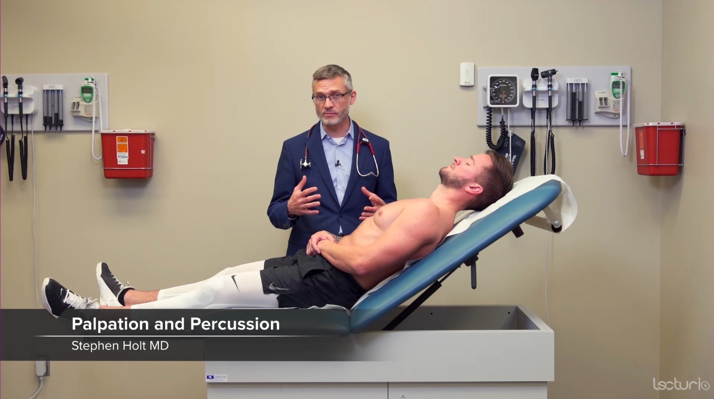
00:01 First off, heart failure you're definitely going to come across patients with acute decompensated heart failure with whether systolic or diastolic or mixed in nature. 00:10 And a few cardinal features can really help us. 00:14 We already talked about the 3rd heart sound, the S3, which again we're going to appreciate with the bell of the stethoscope in the left lateral decubitus position at the apex, but another very important part of the cardiovascular exam when you're trying to assess for heart failure, is to assess volume status and that's best on by looking at central venous pressure as estimated by jugular vein distension, by your jugular vein pressure. 00:40 And the way that we're going to do that is by tilting his head off to the right, just tilt your left, to your left, sorry Shaun. 00:46 We can now visualize the vessels of his great neck in his, underneath his skin. 00:54 So when we're looking at the right side of the neck here, we're trying discern between three different vessels: the carotid artery, the external jugular vein, and the internal jugular vein. 01:04 And there's some very importance features which can help us to distinguish amongst those vessels. 01:08 The carotid artery should have a single way form because it's basically just a systolic impulse, it's causing a jump in the carotid volume and then it's just going to decrease in a very monophasic kind of way. 01:20 The carotid pulsation should also not vacillate, whether he's sitting upright or lying flat. 01:26 It's always going to be pumping at the same volume throughout, and then also, shouldn't be any variation with the respiratory cycle, that is whether he's inhaling or exhaling, the carotid artery will just keep on thumping away in the same pattern in the same volume throughout. 01:43 In contrast, both the EJ, the external jugular and the IJ, the internal jugular will number one, they will fluctuate with the respiratory cycle. 01:53 When I take a deep breath, again I'm creating a vacuum in my body, I'm drawing blood down towards my thorax, so during inhalation, the jugular vein distention should collapse a little bit. 02:05 Likewise, if I lie him flat since this jugular vein is a measure essentially of a column of blood, the amount of pressure that's in his right atrium, if he's standing upright, that column of blood may stop around here at his sternocleido notch, external notch, where as if he is lying flat, then that column of blood is above his horizontal plane so his veins will be completely full. 02:32 So depending on what position I have him in, those veins will either be distended or it will be flat. 02:38 Which is why I've got him right now at about 30 degrees in order to really try and catch the top of that column of blood. 02:46 And the third distinguishing point between jugular veins and the carotid arteries is that it's not going to have a monophasic waveform, its going to have a biphasic waveform. 02:59 That is rather than just going lub-dub, lub-dub, lub-dub kind of picture, I'm trying to keep you timed in with the cardiac cycle, instead it's going to have this kind of picture -- it's one important pulsation at beginning which is actually simply when the right ventricle contracts, it slams close the tricuspid valve and blood backs up a little bit and causes that initial rise, and then the second rise there is basically as the right atrium is now filling up with blood, blood starts to back up again up the superior vena cava and up towards the jugular veins, so you're going to have a biphasic type of appearance. 03:41 So taking a look at Shawn's neck here, you know, some people question whether you can use the external jugular or the internal jugular, I can tell you that you can use both, either one is fine and often times the external jugular, since it is external and more superficial, it's easier to use and so that's what I'm going to use. 04:03 This is his external jugular vein here, there's a little bit of blood distension right here that I can exacerbate in a moment, and this is in contrast to his carotid artery which has a monophasic waveform and that's shown here. 04:21 And his internal jugular which is a bit deeper to the sternocleidomastoid, and it is somewhat more difficult to discern, but let's focus here on the external jugular vein. 04:31 If I push down on that vein and drain the blood from his external jugular vein, down towards his, towards his chest, the external jugular vein in this case is flat. 04:48 If I release my finger you'll see that little swift run of blood from the blood that I was occluding proximally, that is the blood that was draining from his scalp so it's a simple way to make sure you're looking at the right vessel, is to occlude it proximally or cephalad, release the finger then I can see very clearly that vein light up. 05:05 The fact that that vein, the top of it, is right around here, if I follow that plane out here to his external angle, we consider the external angle to mark around 5 centimeters of pressure above the left atrium and he's basically on par with that, maybe one centimeter above that, we can say that his central venous pressure is around 6 cm. 05:25 If in contrast, I'm going to lift you up a little bit Shaun, lie back again. 05:30 If, when I perform this again, if I perform the same maneuver and I found out that that column of blood was up here, if I walk that across parallel to the floor and look at the distance from here down to here which is probably another 5 or 6 centimeters, I would say he has a central venous pressure of 5 plus 5 which is a distance from external notch, down to his left atrium or right atrium, he would have central venous pressure of closer to 10 or 11. 05:58 This is an extremely reliable way to assess central venous pressure and as we'll see when we're looking at pulsus paradoxus and evidence of a pericardial effusion, it's also important to be able to assess these vessels in that context. 06:13 So one strategy to accentuate these jugular veins that you'll see in a person who has heart failure is called the abdominojugular reflux. 06:22 In this case if somebody has too much volume, they're hyperdynamic, they have too much intravascular space, if I squeeze the blood out of his abdomen, I'm going to transiently increase the amount of blood that is regurgitating towards his right atrium and up towards his SVC and ultimately to his IJs where I can see them. 06:43 In a patient who otherwise is well and has good systolic function, I can push on his abdomen, it would be a transient rise, maybe to 1 or 2 or 3 cardiac cycles, but it would very quickly fall back down again, but in a person with heart failure, it'll be sustained, it'll continue to be up there cuz I've pushed all that blood out of his splanchnic vascular bed and its going up towards his jugular veins. 07:07 So if I may, Shaun, let me push right here on your belly, again, while looking at the external jugular vein here and occlude at the top and I'm going to push. 07:27 Now, as my patient does not have any evidence of heart failure on exam, unsurprisingly, I did not see any evidence of abdominojugular reflux. 07:34 When present, it has a very high likelihood ratio in support of volume overload, whether from heart failure or from some other etiology. 07:43 The next two features that would suggest heart failure would be crackles and we're going to defer the respiratory exam to the next lecture, but essentially, looking for crackles can support the diagnosis of heart failure. 07:58 We'll keep in mind that crackles are so non-specific that they don't really have a significant likelihood ratio, whether positive or negative, in support of heart failure. Instead, they're very useful to track a patient from one day to the next, while you're trying to diurese them and decrease their volume. 08:15 And then the last one is peripheral edema, so let's go take a look down in this legs and see if we can investigate that.
About the Lecture
The lecture Intravascular Volume Assessment – Findings in Heart Failure by Stephen Holt, MD, MS is from the course Examination of Cardiovascular and Respiratory System.
Included Quiz Questions
What is TRUE regarding abdominojugular reflux?
- Abdominojugular reflux is due to an increased inflow of blood from the abdominal veins into the right atrium.
- Abdominojugular reflux is a prolonged increase in jugular venous pressure seen in patients with dehydration.
- Abdominojugular reflux is a physical examination test useful in diagnosing emphysema.
- Abdominojugular reflux is normally about 10 cm above the sternal notch.
- Abdominojugular reflux is a reflection of the right ventricle compensating for augmented venous return.
An elevated central venous pressure reflects...
- ...volume overload.
- ...elevated blood pressure.
- ...normal compensation with heart failure.
- ...the monophasic venous pulse.
- ...the carotid arterial impulse.
The venous pulse found on physical exam of the neck is...
- ...biphasic, easily obliterated by light pressure, and increased in the supine position.
- ...monophasic, easily obliterated by light pressure, and decreased in the supine position.
- ...monophasic, not obliterated by light pressure, and increased in the supine position.
- ...biphasic, not obliterated by light pressure, and decreased in the supine position.
- ...biphasic due to left atrial and right atrial contractions.
Physical exam findings seen in heart failure include...
- ...increased jugular venous pressure, positive abdominojugular reflux test, and peripheral edema.
- ...decreased jugular venous pressure, positive abdominojugular reflux test, and crackles in the lungs.
- ...decreased jugular venous pressure, negative abdominojugular reflux test, and crackles in the lungs.
- ...increased jugular venous pressure, negative abdominojugular reflux test, and poor skin turgor.
- ...increased jugular venous pressure, the point of maximal impulse displaced to the left, and jaundice.
Author of lecture Intravascular Volume Assessment – Findings in Heart Failure

Stephen Holt, MD, MS
Customer reviews
5,0 of 5 stars
| 5 Stars |
|
1 |
| 4 Stars |
|
0 |
| 3 Stars |
|
0 |
| 2 Stars |
|
0 |
| 1 Star |
|
0 |
Great review! I appreciate how easy it is to follow!
