Playlist
Show Playlist
Hide Playlist
Intrahepatic Biliary System

-
Slides Gallbladder and Spleen.pdf
-
Download Lecture Overview
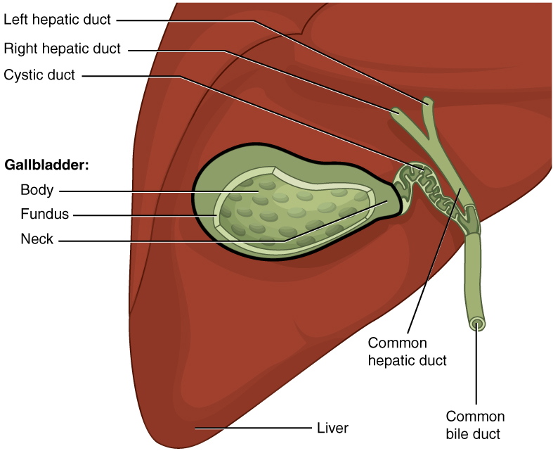
00:00 So let's review the intrahepatic biliary system a little bit as well. 00:04 The intrahepatic bile duct should really be less than about 3 millimeters or so in diameter. Most commonly this can be dilated due to choledocholithiasis or obstruction of the common bile duct with a stone or another abnormality such as sludge. 00:18 They can become dilated due to any kind mass lesion as well which obscures the central biliary system. 00:24 Patients that have a central obstructing lesion often present with jaundice and pruritis. 00:30 So those are the two most common findings of a patient presenting with a central obstructing lesion. 00:35 So this is an example of intrahepatic biliary ductal dilatation. 00:41 We can see here two ultrasound images, so here we have what look like dilated ducts. 00:48 You can see that on the Doppler you can tell which one is the portal vein versus which one is a bile duct. 00:54 Although we had mentioned ways of telling the difference where the portal vein has an echogenic rim around it and the bile duct doesn't, sometimes it can be difficult especially when the bile ducts are dilated so the other way of telling is to place Doppler and to see which one fills with color. 01:06 The one that fills with color is going to be the vessel and the one that doesn't is going to be the duct. 01:11 So the duct isn't dilated here and if you look at the axial CT image, you can see contrast within the portal venous system and then surrounding the portal venous system we see linear areas that are hypodense and these represent dilated bile ducts. 01:25 So what is an MRCP? It stands for Magnetic Resonance Cholangiopancreatography. 01:32 It's a heavily T2 weighted sequence that's performed without contrast and it's actually very helpful in determining the cause and level of obstruction of the biliary system. 01:41 So this is an example of an MRCP. 01:46 You can see that there's mild intrahepatic and extrahepatic biliary ductal dilatation so these here represent intrahepatic bile ducts and then as you come more centrally you can see dilated extrahepatic bile ducts as well. 01:58 The extrahepatic ducts abruptly end right here at the level of an obstruction. 02:03 You can see here a similar MRCP image which shows hyperintens and then an abrupt end where you see this dark round structure and this represents a stone that's obstructing the distal common bile duct. 02:15 So an MRCP is a very good substitute to an ERCP an ERCP is performed by a GI physician and it's a lot more invasive than an MRCP is so often we start with an MRCP first to take a look at the level of obstruction before we proceed to an ERCP which can help resolve the obstruction as well. 02:32 So cholangiocarcinoma, this is a very important malignancy of the biliary system. 02:36 It can be located really at any level of the biliary tree and the most common is the Klatskin tumor which is cholangiocarcinoma located at the confluence of the right and left hepatic ducts. 02:47 So here is an example of a cholangiocarcinoma. 02:51 We can see two CT images. 02:53 We have an axial CT image, we have a coronal image, and then we have again an MRCP image. 02:59 And you can see that there's a low density mass at the confluence of the right and left hepatic ducts and this results in an abrupt cut off of the biliary system as you can see by this arrow here. 03:09 So we have multiple intrahepatic ducts that look dilated and then we have an abrupt cut off when we actually don't see the extrahepatic biliary system because of this mass that's obstructing the biliary system and this is an example of what a cholangiocarcinoma would look like.
About the Lecture
The lecture Intrahepatic Biliary System by Hetal Verma, MD is from the course Abdominal Radiology.
Included Quiz Questions
A patient presents with right upper quadrant pain and has an ultrasound demonstrating intrahepatic and extrahepatic biliary ductal dilatation. What is the next step to determine the cause?
- MRCP
- Abdominal CT with contrast
- Abdominal CT without contrast to evaluate for stones
- HIDA scan
- No further imaging is needed
Which of the following is TRUE regarding cholangiocarcinoma?
- Cholangiocarcinoma can be located at any level of the biliary tree.
- A Klatskin tumor is cholangiocarcinoma located at end of the left hepatic ducts.
- A CT scan will show a Klatskin tumor as a well-defined mass at the confluence of the middle and main hepatic ducts.
- There is an abrupt cutoff of the bile ducts on imaging of Klatskin tumor.
- Cholangiocarcinoma is a metastasis of hepatic cancer into the biliary system.
Author of lecture Intrahepatic Biliary System

Hetal Verma, MD
Customer reviews
5,0 of 5 stars
| 5 Stars |
|
5 |
| 4 Stars |
|
0 |
| 3 Stars |
|
0 |
| 2 Stars |
|
0 |
| 1 Star |
|
0 |
