Playlist
Show Playlist
Hide Playlist
Hypertensive Retinopathy

-
03 Retinal Disorders V2.pdf
-
Download Lecture Overview
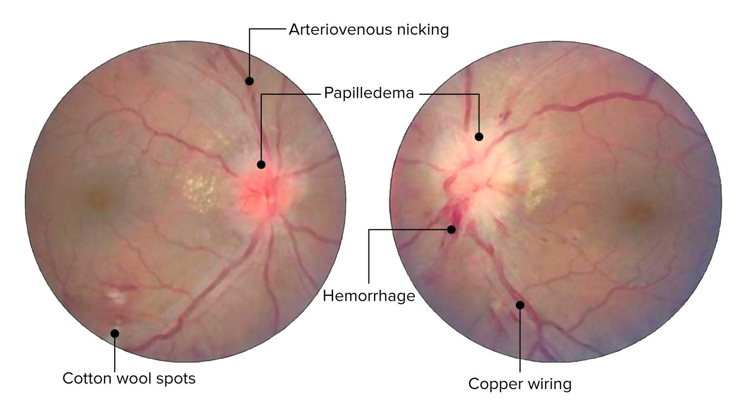
00:01 Along with diabetes, a patient often times in the US will have hypertension, correct? They go hand in hand, unfortunately. 00:07 Now when you have hypertension in your blood vessel then you could undergo, let's say benign hypertension, which is what, 95% of your patients. 00:16 So, on three different office visits, the blood pressure has been 140/90, right? So now the arterials will undergo changes that we call hyaline atherosclerosis. 00:30 What's happening here is there's accumulation of protein. 00:34 In the walls of your blood vessel causing it to become narrowed and may result in, once again, dot-blot hemorrhages or cartwheel spots, keep that in mind. 00:42 Or let's say that you have an African-American male, of approximate 25 years of age, and has a blood pressure of 212/180, that's malignant hypertension. 00:56 And therefore now the blood vessels in the retina may undergo hyperplastic arteriosclerosis. 01:02 The description of that is then called onion skinning, is that clear? Here's a picture of onion skinning, secondary to malignant hypertension in which the arrow here is then pointing to areas in which there is smooth muscle hyperplasia. 01:21 This is hyperplastic arteriolosclerosis, secondary to malignant hypertension. 01:27 Giving rise to in the eye, in the retina, hypertensive retinopathy. 01:32 We have something called the modified Scheie Grade or grades. 01:37 And quickly here, It's not that, that it's important that you pay attention to every single grade, but keep in mind the relative destruction that's taking place as we go from grade one, in which for the most part, there's mild narrowing of your retinal blood vessels. 01:53 And you go into grade two in which, here once again, compression of the retinal veins; so what's gonna happen when you have retinal vein compression? In fact, later on I'm gonna give you retinal vein occlusion. 02:07 You can no longer drain into your optic disc, and so therefore, you're gonna accumulate blood back in, and that particular blood vessel is going to be engorged. 02:17 And you go into grade three, in which what happens now is the fact that you have your bleeding and hemorrhage taking place. 02:25 There might be sclerosis which we then called, 'silver wire,' just like the name implies that means sclerosis. 02:33 Sclerosis means thickening and we call this in pathology, silver wire appearance. 02:40 And if it gets really bad in terms of your blood pressure, and let's say that you go into states of let's say, malignant hypertension. 02:47 Then you might have distortion of the optic disc, and when that happens, we call this papilledema. 02:54 Better keep that in mind. 02:55 This is going to choke everything within the disc, extensive hemorrhage. 02:59 Not a good thing. 03:01 That's a Grade IV. 03:02 Here, we'll take a look at central retinal artery occlusion. 03:06 Picture that for me first. 03:07 So that would be the retinal artery that is coming out of the optic disc, isn't it? Yes. 03:13 Along with the central vein which is bringing, reigning blood in, and you have the optic nerve, which obviously is taking in impulses. 03:20 You're gonna look for a patient. 03:23 Who, in which there's sudden, painless, complete loss of vision in one eye. Okay? Monocular. That's important. 03:33 Painless, sudden. 03:35 How this occur? Maybe there was embolization. 03:39 When there is embolization that's taking place there might be every possibility as central retinal artery may then become occluded. 03:48 What you're gonna notice here, pallor of the optic disc due to the narrowed artery, exact what we are seeing here, on this fundoscopic examination, and the optic disc is a lot more pale and the blood vessel specific at central retinal artery, coming out of there is going to be occluded. 04:05 Look for sudden, painless, complete loss of vision in one eye. 04:09 Please, that's central retinal artery occlusion, secondary to embolization. 04:13 Embolization, coming from? Maybe the carotid, ophthalmic artery plaque, or perhaps even, remember, when you have a, let's say, an older patient that has pain, maybe up here in the temporal region in the jaw. 04:28 And you do a biopsy, in the end you find the granulomatous type of inflammation of the temporal artery. 04:33 If left untreated, remember the ophthalmic branches, and such? May then give you cause of blindness, but could be secondary to Giant-cell arteritis as well. 04:43 Keep that in mind. 04:44 Or more commonly embolization. 04:47 Central retinal artery occlusion. 04:49 Compare that to what's happening here. 04:52 Central retinal vein occlusion. 04:55 This also will be sudden and painless, also monocular, but this time, take look at that fundoscopic examination. 05:04 A lot more engorgement, of your blood vessel. Why? Because the central, in general, vein is responsible for draining. 05:11 To any vein that becomes occluded, what's gonna happen proximally? Engorgement, congestion. This is congestion, isn't it? That's central retinal vein occlusion. 05:23 For example, when you think about testicular torsion, take a look at that knot, then take a look at the scrotum. 05:28 It's red, isn't it? Really dark. 05:30 Because here when the spermatic cord becomes occluded, the vein also then becomes occluded, and this congestion taking place in the scrotum. 05:38 Here take a look, there's congestion. 05:41 So it's engorged retinal vein is what you're focusing upon. 05:44 We call this, blood and thunder appearance, it looks a heck of a lot more dangerous and significant than the previous picture, which was the central retinal artery occlusion. 05:57 Now, it's due to? Good. 06:00 Embolization most commonly. 06:02 Here we have congestion taking place.
About the Lecture
The lecture Hypertensive Retinopathy by Carlo Raj, MD is from the course Retinal Disorders. It contains the following chapters:
- Hypertensive Retinopathy
- Central Retinal Artery Occlusion
Included Quiz Questions
Which type of hypertension most likely leads to onion-skinning of the blood vessels on histology?
- Malignant hypertension
- Stage I hypertension
- Stage II hypertension
- Primary hypertension
- Secondary hypertension
Papilledema occurs in which grade of hypertensive retinopathy?
- Grade IV
- Grade I
- Grade II
- Grade III
- Grade 0
A "blood and thunder" appearance on fundoscopic examination of a patient with sudden, painless, unilateral loss of visual acuity makes which diagnosis most likely?
- Central retinal vein occlusion
- Central retinal artery occlusion
- Open-angle glaucoma
- Closed-angle glaucoma
- Retinal detachment
What is the most common cause of central retinal artery occlusion?
- Embolism
- Vasospasm
- Giant cell arteritis
- Dissecting aneurysm
- Idiopathic
Author of lecture Hypertensive Retinopathy

Carlo Raj, MD
Customer reviews
5,0 of 5 stars
| 5 Stars |
|
5 |
| 4 Stars |
|
0 |
| 3 Stars |
|
0 |
| 2 Stars |
|
0 |
| 1 Star |
|
0 |
