Playlist
Show Playlist
Hide Playlist
How Are Connective Tissues Classified?

-
Slides 03 Types of Tissues Meyer.pdf
-
Reference List Histology.pdf
-
Download Lecture Overview
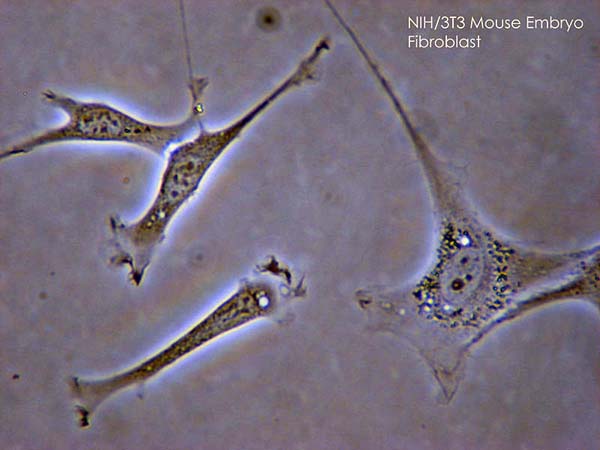
00:02 Well, it is important that we start off by learning something about how connective tissue is classified, how it is named. All connective tissue arises in the embryo from mesenchyme or mesoderm. 00:22 Sometimes that mesenchyme differentiates into what we call mucous connective tissue in the umbilical cord and I will talk about that later on during this lecture. But mesenchyme developing into the mesoderm then provides all the necessary cells and components of connective tissues. We will learn later on that mesenchyme also gives rise to smooth muscle and muscle to do with the cardiovascular system and the urogenital system and serous membrane. So mesenchyme is not just restricted to providing connective tissue. Now, in the adult, you can divide connective tissue into three basic components or three basic levels of classification. The most common one is just ordinary connective tissue such as lamina propria, which is loose connective tissue or dense irregular connective tissue like the dermis of skin or regular connective tissue like a tendon. Sometimes we classify connective tissues as being supporting such as cartilage and bone. And there are also very special connective tissues, adipose tissue, which stores fat, stores energy, reticular tissue, we are going to talk about during our talks on the lymphatic system and of course blood as well. 02:01 Here is an image of a developing part of the body. It is embryonic. And the reason why I want you to have a look at this very carefully is because I want to show you a mesenchyme cell. 02:14 A mesenchyme cell is a very very thin elongated nucleus and it consists of cytoplasm that we cannot see because that cytoplasm is spread out throughout the connective tissue, very very thin. All we see a very spindle-shaped nucleus. Now that mesenchyme cell is going to give rise to all the connective tissue cells that we will learn about. In this case, it is an undifferentiated cell. But during development, as we see here, it can be told to develop into a tissue. Here that mesenchyme cell is being told, you will form bone. So that mesenchyme cell starts to differentiate and form an osteoblast. And here we see some osteoblasts lying on the surface of bone that has been created. Those osteoblasts are lying down bone matrix and you see lots of little dark nuclei belonging to these osteoblasts and they are sitting on top of the surface of newly formed bone. And when those bone cells of secreted matrix they surround themselves and then they sit there and maintain that matrix as osteocytes. And we will talk more about bone development, bone growth, bone formation and the role of these osteoblasts and osteocytes in a later lecture. But the key point is mesenchyme cells are undifferentiated and they can be told to differentiate further into the osteoblast and then other connective tissue cells as well. 04:07 Here is a slide, a summary of all the sorts of cells in connective tissue that we will cover in this lecture and in a subsequent lecture on cartilage and bone. 04:21 A fibroblast, a mesothelial cell, endothelial cells that line blood vessels and also chondroblasts and chondrocytes that form a cartilage and as we saw previously, the osteoblast and the osteocytes in bone. 04:40 Let's look at some of these classifications of connective tissue in a little bit more detail. Here is a section through loose connective tissue or areolar connective tissue. 04:53 You know it is called areolar connective tissue because in the very early days, when the early anatomists and histologists tried to describe these connective tissues, when they peel the skin away from an animal, the gap between the dermis and the hypodermis accumulate a little air bubbles. 05:12 So they called it is aerated connective tissue or areolar tissue. That is where it gets its name from. But have a look at this picture, you can see at the very top, some rather dense stained connective tissue, collagen. It happens to be the wall of a blood vessel, but underneath you can see very sparse cells wandering through this loose connective tissue. That clear space dominates and that clear space is the ground substance. Sometimes you can see fine little collagen fibres running through this particular connective tissue. So this is loose connective tissue such as we saw in the lamina propria. Here is the lamina propria again just to remind you of what it looks like. It supports the epithelium, the basement membrane is the cement or the structure that holds that epithelium bound to the underlying lamina propria or the connective tissue. And if you look very carefully in this slide, in the lamina propria, you can make out some very fine little red dots and red fibres. These are going to be both collagen and elastic tissue that we will talk about later on. This gives the lamina propria support, structure and strength, cohesion, but it also gives the lamina propria flexibility because epithelial serves moves. They are mobile and they are only mobile because of the looseness of the underlying connective tissue and the components of the fibres within that lamina propria. Here is another example of areolar tissue or loose connective tissue. 07:07 Here is a sweat gland shown here and just around the sweat gland, you have very loose pieces of connective tissue. This is now dense irregular connective tissue. Notice how different it is to what you saw previously with the lamina propria. The nuclei you see here are mainly nuclei belonging to either endothelial cells of capillaries going to the surface of the skin, this happens to be the epidermis sitting on the dermis. Or they're the nuclei of may be some fibroblasts that the collagen here dominates. You can see collagen that sectioned longitudinally and you can say collagen that section transversely. Now this provides a dermis with enormous strength because the collagen is arranged in lots of different networks and in lots of different directions that prevents the skin from tearing. It gives the skin strength and also some mobility. The epidermis is cemented directly onto this dermis. Now there is are too many cells there to do with the immune system and yet skin is an epithelium. 08:30 It is a very specialised epithelium and that is because the epidermis that is labelled here has special components that prevent pathogens from invading through the epidermis into the underlying dermis. The waterproofing layer of the keratin, the lipid and the protein layers that are insoluble that are on the outside and inside of the membrane of the stratum corneum, and the Langerhans cells or the antigen presenting cells within the epidermis are all there to be a barrier to invading pathogens. So in the case of dermis, you do not see the sorts of immune cells or presence of lots of nuclei that are under surveillance or sentry duty as you do in the lamina propria underneath an epithelium. 09:25 Here is an example of dense regular connective tissue such as the tendon. Here the collagen bundles are arranged in parallel. As I mentioned earlier, the job of a tendon is to impart contraction from a muscle directly onto the bone. So to do that effectively, the collagen is arranged parallel. And if you look very very carefully, you can see fibrocytes. 09:53 These are fibroblasts that they have done their job laying down the fibres so they are resting and undergoing just maintenance of the collagen. The resting fibroblasts we often term a fibrocyte. 10:10 Now in the case of a tendon, these cells can get another name. They are called a tendinocyte. 10:18 And these collagen bundles, if we can see the outside surface of the collagen, there would be a very thin capsule around the collagen bundles or around the tendon, and the collagen in this capsule would be arranged less sort of parallel than you see here. And that capsule is called the epitendineum. Each bundle of collagen is wrapped up by endotendineum and we will come across terminology like that when we describe the wrappings up of muscle cells and also nerve cells.
About the Lecture
The lecture How Are Connective Tissues Classified? by Geoffrey Meyer, PhD is from the course Connective Tissue.
Included Quiz Questions
Which of the following best describes the composition of the reticular dermis?
- Dense irregular connective tissue
- Loose regular connective tissue
- Dense regular connective tissue
- Loose irregular connective tissue
Which of the following is the main embryologic origin of connective tissue?
- Mesoderm
- Endoderm
- Ectoderm
- Yolk sac
- Notochord
Which of the following is NOT a type of connective tissue?
- Epithelium
- Adipose tissue
- Blood
- Bone
- Cartilage
Author of lecture How Are Connective Tissues Classified?

Geoffrey Meyer, PhD
Customer reviews
4,0 of 5 stars
| 5 Stars |
|
1 |
| 4 Stars |
|
0 |
| 3 Stars |
|
1 |
| 2 Stars |
|
0 |
| 1 Star |
|
0 |
Outstanding review of connective tissue! These videos are helping me get so much more out of my studies!
CT proper: mesenchyme, lax fibrillary, dense fibrillary, reticular, elastic, adipose and pigment. Supporting CT: cartilage and bone. Trophic CT: blood and lymph.
