Playlist
Show Playlist
Hide Playlist
Heart: Summary
-
Slides 01 Human Organ Systems Meyer.pdf
-
Reference List Histology.pdf
-
Download Lecture Overview
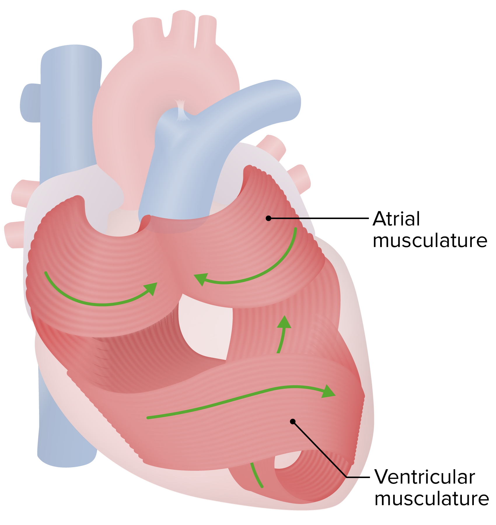
00:00 So, in summary, endothelium is a very important epithelium because it lines the entire vascular system. And as we go through other lectures in this histology course, I will emphasize all the different sorts of functions that endothelium has. Recall that the atria of the heart receive blood, from the systemic circulation or from the lungs. The ventricles are the pump of the heart that sends blood to the lungs to be oxygenated, and then it’s returned to the left side of the heart where it’s pumped by the left ventricle to the systemic circulation. And the wall has got three distinct layers: the outside layer, the epicardium, the inner myocardium, which is where all the pumping action occurs because that’s composed mostly of cardiac muscle. And then an internal lining, the endocardium, which includes the endothelium supported by some connective tissue underlying that epithelial layer. Cardiac muscle has only got one nucleus. It’s striated. 01:24 It’s branched. It’s arranged and joined end to end with other cardiac muscle fibres by intercalated discs which contain adherent junctions to make sure they’re bound very tightly together. 01:39 But also they contain gap junctions to enable transmitters, messengers, ions, to go from one cell to the other, and importantly, bring about a sequential contraction of cardiac muscle. The heart pumps because impulses are generated at the sinoatrial node. And then they are carried across, from the atria across a fibrous skeleton to the ventricles, where they initiate contraction again of the ventricles. And that initiation of contraction of the ventricles is acted upon by the presence of Purkinje fibres. These Purkinje cells transmit the impulse through the myocardium to all the cardiac muscle cells within that myocardium. 02:34 And it’s important to know the difference between the cardiac muscle fibres that contract and these Purkinje fibres that just transmit the impulse. Recall that the Purkinje fibres, being just a conducting muscle cell, don’t contract, and therefore, don’t have the apparatus to contract. Therefore, they don’t stain as heavily as normal cardiac muscle cells stain. So I hope you’ve enjoyed this lecture. I hope you now have a good understanding of the histological structure of the heart and the importance of the heart as being a component of the cardiovascular system. Thank you.
About the Lecture
The lecture Heart: Summary by Geoffrey Meyer, PhD is from the course Cardiovascular Histology.
Included Quiz Questions
Which of the following statements regarding the Purkinje fibers is NOT correct?
- They have contractile properties.
- They propagate an action potential.
- They contain pale staining with hematoxylin and eosin.
- They are present in the ventricles.
Customer reviews
5,0 of 5 stars
| 5 Stars |
|
3 |
| 4 Stars |
|
0 |
| 3 Stars |
|
0 |
| 2 Stars |
|
0 |
| 1 Star |
|
0 |
i really enjoyed the lecture, it was very clear and easy to understand.
This is really good, and easy to understand, thank you.
?ts so d?rect clear and understanble...one of best h?stology lectures so far


