Playlist
Show Playlist
Hide Playlist
Gluteal Region


-
Slide Gluteal Region.pdf
-
Reference List Anatomy.pdf
-
Download Lecture Overview
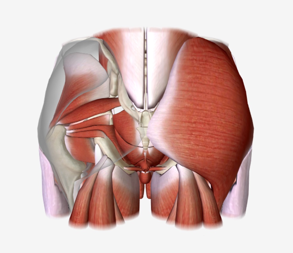
00:01 So in this lecture, we're going to look at the gluteal region And it starts off with, let's just introduce ourselves to where these are located within the lower limb. 00:11 So here we can see the posterior aspect of the right lower limb. 00:16 And we can see we have indicated here, the gluteal region, most proximately, or most superiorly, and then we have the posterior thigh and then popliteal region. 00:26 So these key regions that we're going to discuss is part of the lower limb. 00:30 So lets move on to the gluteal region. 00:33 So the gluteal region really is a mass of muscles that form the buttock and here we're looking at the postural lateral aspect of this right gluteal region. 00:45 Here we can see it's made up of a whole series of large broad muscles, we have gluteus Maximus, we have gluteus medius. 00:53 And then deep to gluteus medius, we have gluteus minimus. 00:57 And then most laterally, we have the muscle tensor fascia lata. 01:02 These muscles formed very much a superficial group within this gluteal region. 01:07 Later on, we'll see some deeper muscles that have also residing within this area. 01:13 The iliotibial tract is an important fascial continuation. 01:17 It's connected to the tensor fascia lata muscle, and it helps to stabilize both the hip joint and the lateral aspect of the knee joint. 01:25 It's continuous with the deep fascia of the thigh. 01:29 Now let's have a look at some of these muscles in more detail. 01:32 We've got gluteus Maximus, we can see on the screen at the moment. 01:36 And its origin comes from the ilium, we can see here, it also comes from the sacrum, it comes from the coccyx, and it comes from the sacrotuberous ligament. 01:46 So it's origin forms this C-shaped attachment site, we can see there ilium, sacrum, coccyx, and the sacral tuberous ligament. 01:55 It then extends laterally away to form the iliotibial tract, and it attaches to the gluteal tuberosity, which we can see here on the postural lateral aspect of the femur. 02:09 It's applied by the inferior gluteal nerve which we can see here. 02:13 Remember, the inferior gluteal nerve is passing out underneath piriformis. 02:18 So the function of gluteus Maximus can be seen here, we have hip extension with the femur moving posteriorly within the sagittal plane, and then also it allows the hip to laterally rotate, so move the femur laterally in that rotational direction. 02:35 So now let's move on to gluteus medius. 02:37 It's in a similar region but a much smaller muscle. 02:40 We can see the origin of gluteus medius is here coming away from the ilium and we see a passes towards the greater trochanter of the femur. 02:49 This muscle is innervated by the superior gluteal nerve, which emerges out of the greater sciatic foramen running superior to piriformis. 02:58 The function of gluteus medius is to help medially rotate the hip joint and it also because it's location helps to abduct the hip. 03:06 So move the hip or move the femur at the hip joint away from the midline of the body. 03:14 Now let's look at gluteus minimus. 03:16 Gluteus minimus is the smallest of these three gluteal muscles. 03:19 It again comes from the external surface of the ilium which we can see here. 03:24 And it passes also to the greater trochanter of the femur. 03:29 It is also like gluteus medius supplied by the superior gluteal nerve. 03:34 The function of gluteus minimus is again, to help medially rotate the hip joint and it also helps to abduct the hip as well. 03:42 So abducts the femur at the hip joint moves the femur away from the midline. 03:48 Now if we look at the final muscle in this superficial gluteal region collection, we have tensor fascia lata muscle. 03:55 Here we can see the origin of the tensor fascia lata muscle is from the anterior superior iliac spine. 04:01 And then you see actually blends with this dense connective tissue fascial tract known as the iliotibial band, and that runs all the way down and attaches to the lateral condyle of the tibia. 04:14 So this one doesn't directly insert into a muscle similarly to the other gluteal muscles, it very much forms that connective tissue band, which ultimately then runs to the lateral condyle of the tibia. 04:25 Innervation of this muscle is why the superior gluteal nerve and the function is very much to help flex the hip alongside other muscles, but importantly helps us stabilize the knee joint as it's running along the lateral aspect. 04:39 And that's important how to maintain posture. 04:44 So now let's continue looking at muscles within the gluteal region. 04:47 Here we're going to look at a series of muscles, here's piriformis, superior gemellus, obturator internus, and inferior gemellus. 04:57 The final muscle here is quadratus femoris. 05:00 So what we're doing is we're looking at the postural lateral view of the gluteal region, we're on the right hand side and see a series of muscles that are lying deep to those gluteal muscles we just described. 05:12 The final muscle we can see here, and it's a slightly complicated muscle is obuturator externus. 05:17 So that's quite hard to see. 05:19 But we'll pick that up as we move forward. 05:22 These muscles are known as the deep muscle group, and like I said, they sit deep to the gluteal muscles. 05:28 Let's look at these individually one by one. 05:31 Here we have piriformis muscle. 05:33 Piriformis muscle actually emerges from the anterior surface of the sacrum. 05:37 So it's coming away from the anterior surface of the sacrum within the pelvis, but it exits the pelvis via the greater sciatic foramen. 05:46 And as it does this, it passes laterally towards the femur. 05:50 And we can see here it attaches to the greater trochanter of the femur. 05:55 We also have a muscle that sits inferior to it. 05:58 This is the superior gemellus muscle, it originates from the ischial of spine on the ischium, and also passes towards the greater trochanter. 06:06 Specifically, the more medial aspect to it, because piriformis is occupying a more superior position. 06:14 We then have the obturator internus muscle, this muscle is also coming from inside the pelvis, it's coming from the internal surface of the obturator membrane. 06:25 So here we can see coming from the obturator membrane on the inside of the pelvis, and also the surrounding bones. 06:31 To remind ourselves of those bones that form the obturator foramen, please do look at the bone lecture, but it's coming out of the pelvis through the lesser sciatic foramen, and it passes over into the gluteal region and attaches to the greater trochanter of the femur. 06:51 Here we can see the inferior gemellus. 06:52 And this inferior gemellus is coming from the ischial tuberosity. 06:56 And that also passes to the greater trochanter. 06:59 So now let's look at the innervation of these muscles, we can see we have a series of nerves that really have named branches. 07:07 So here we have a nerve to piriformis. 07:09 It comes from S1, S2. 07:11 And then the nerve to obturator internus is supplying the superior gemellus and obturator internus muscle. 07:18 It also may supply inferior gemellus. 07:20 But sometimes we have nerve to quadratus femoris which we'll see in a moment that can provide innervation to this muscle. 07:27 Essentially, we have some branches that are coming directly from the sacral plexus and these are supplying the shorts muscles around the hip joint. 07:37 If we look at the function of these muscles, then we can see actually they are all together responsible for lateral rotation of the extended hip. 07:46 And they also help because of their position on the greater trochanter, they help to abduct the hip as well. 07:52 So lateral rotation of an extended hip abduction of a flexed hip. 07:57 So now let's talk about quadratus femoris. 08:00 Here we can see Quadratus Femoris, it's coming away from the ischial tuberosity. 08:04 And it's passing laterally to the intertrochanteric crest. 08:09 So it's passing laterally from the ischial tuberosity to the intertrochanteric crest. 08:15 That crest on the posterior surface between the greater and lesser trochanter. 08:19 It's applied by nerve to Quadratus Femoris. 08:22 It's responsible for laterally rotating the extended hip. 08:28 Let's also have a look at obturator externus. 08:30 So this muscle is slightly difficult to see and we've removed all the other muscles so we can visualize it. 08:36 But whereas obturator internus was coming from the arbitrator membrane and surrounding muscles on the inside of the pelvis. 08:45 Obturator externus is doing the opposite but from the external surface. 08:51 So it's coming from the external surface of obturator membrane and the surrounding bones or the anterior surface. 08:57 This muscle also passes laterally and it attaches to the trochanteric fossa of the femur. 09:05 This muscle is innervated by the obturator nerve, and it's responsible for laterally rotating the extended hip. 09:13 So we have a series of muscles here that are responsible for both the medial rotation and the lateral rotation of the hip. 09:19 Depending on their origin and insertion that will determine if they can either immediately or laterally rotate the hip joint.
About the Lecture
The lecture Gluteal Region by James Pickering, PhD is from the course Anatomy of the Gluteal Region.
Included Quiz Questions
What is the most lateral muscle of the gluteal region?
- Tensor fasciae latae
- Gluteus medius
- Gluteus minimus
- Gluteus maximus
- Piriformis
What is the origin of the gluteus maximus?
- Sacrum
- Gluteal tuberosity
- Iliotibial tract
- Greater trochanter
What are the functions of the gluteus maximus? Select all that apply.
- Hip extension
- Lateral rotation of hip
- Hip flexion
- Hip abduction
- Medial rotation of hip
What is the function of the gluteus medius?
- Medial rotation of hip
- Lateral rotation of hip
- Hip extension
- Hip flexion
What is the function of the tensor fasciae latae? Select all that apply.
- Hip flexion
- Knee joint stabilization
- Foot extension
- Hip adduction
- Hip torsion
Author of lecture Gluteal Region

James Pickering, PhD
Customer reviews
5,0 of 5 stars
| 5 Stars |
|
5 |
| 4 Stars |
|
0 |
| 3 Stars |
|
0 |
| 2 Stars |
|
0 |
| 1 Star |
|
0 |
