Playlist
Show Playlist
Hide Playlist
Epithelium: Surfaces of Epithelial Cells

-
Slides 01 Types of Tissues Meyer.pdf
-
Reference List Histology.pdf
-
Download Lecture Overview
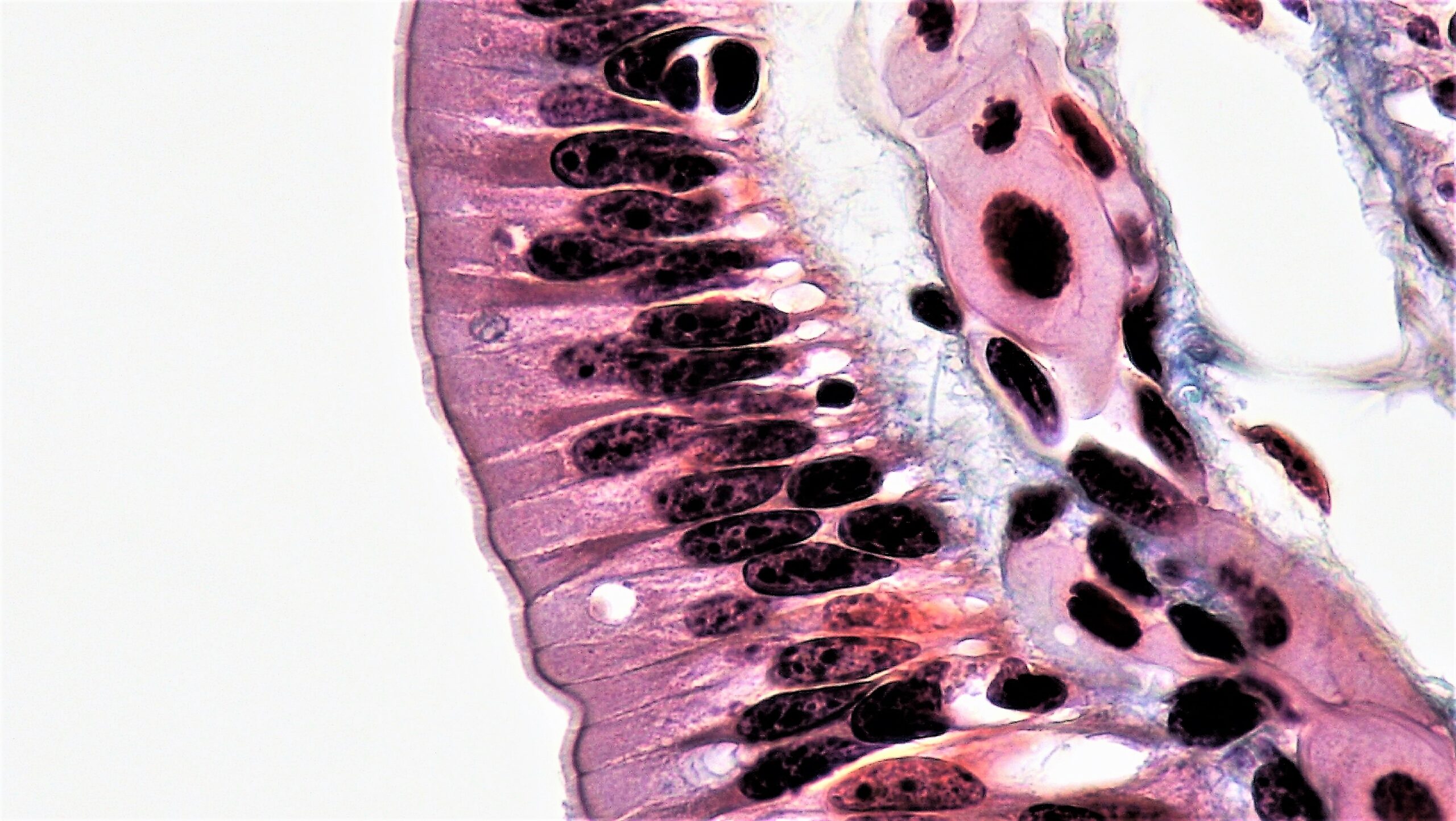
00:01 The lateral surfaces of epithelial cells also are very specialized. In this electron micrograph, there are interdigitizations between projections of adjacent cells. These interdigitizations are very important because they allow the distance or the space between cells to actually expand and this is important sometimes in the transport of fluid. Normally, sodium pumps pump sodium into the space between cells in these lateral border regions and chloride ions and or anionic substances follow to maintain electrochemical stability. And that immediately creates an osmotic gradient. So water passes then from the cell into these lateral regions. 01:03 And on the other image you see the section through an epithelium of the gallbladder, you can see white clear spaces between the cells. This is an example of where water is passing from the cell into these lateral spaces and then the occluding junctions prevent that water from then going to the surface. The hydrostatic pressure builds up and so the water flows back into the connective tissue underneath the cells and then to be absorbed then by the body. And this is the way of the gallbadder being able to concentrate bile. The basal surface of cells also are very specalized. Here you see on the left hand side a number of mitochondria aligned along the basal surface of epithelial cells. There are also lots and lots of basal folds of the cell membrane. And that's because across this epithelial surface, there is a lot of transport of fluid as well and other substances. So the mitochondria there to provide the energy for active transport, and all the foldings is to increase the surface area for these transport proteins and transport channels. And when you look at the sections of epithelia that have these sorts of specializations, they have a rather striated border. Little stripes appear at the basal part of the cells that we call the striated border. And these are typical of striated ducts in protein secreting glands that I will talk about when we look at glands in our later lecture. There they are there shown on the image, very fine pink striations.
About the Lecture
The lecture Epithelium: Surfaces of Epithelial Cells by Geoffrey Meyer, PhD is from the course Epithelial Tissue.
Included Quiz Questions
Which of the following is NOT a characteristic of the basal epithelial cell surface?
- The basal cell surface uses osmotic gradients to allow water accumulation between neighboring cells.
- The basal surface is the surface of the cell adjacent to the basement membrane.
- Various transport proteins and channels are found embedded in the membrane surface.
- Mitochondria in this area are used to generate energy for active transport.
- They often have a folded structural arrangement to increase surface area.
Author of lecture Epithelium: Surfaces of Epithelial Cells

Geoffrey Meyer, PhD
Customer reviews
1,5 of 5 stars
| 5 Stars |
|
0 |
| 4 Stars |
|
0 |
| 3 Stars |
|
0 |
| 2 Stars |
|
1 |
| 1 Star |
|
1 |
Dr. Meyer does not explain where in the illustration he is referring to. It was very hard to follow through.
lecture should be more simplify as it is many times difficult to understand .
