Playlist
Show Playlist
Hide Playlist
Epithelium: Classification

-
Slides 01 Types of Tissues Meyer.pdf
-
Reference List Histology.pdf
-
Download Lecture Overview
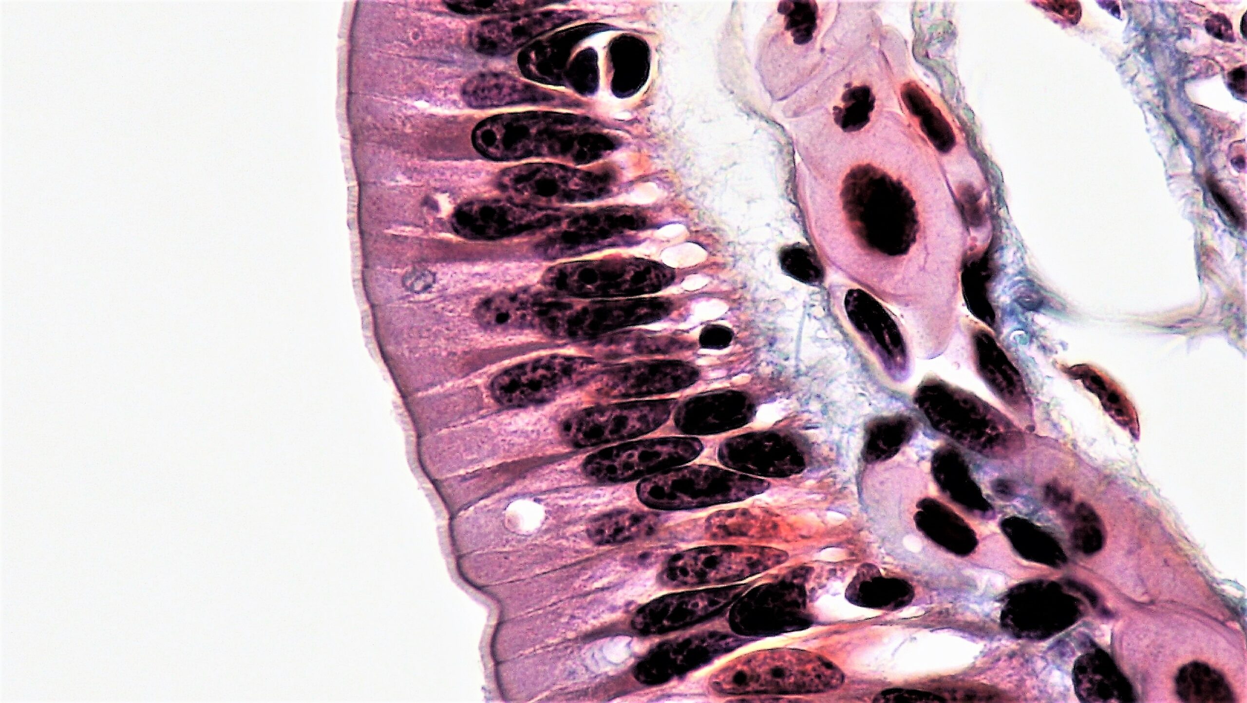
00:01 Let us now look and see how epithelium is classified or named. Well, the first thing to do when you look at an epithelium is decide how many cell layers make up the epithelium. If there is only cell layer as you see in these diagrams, we call the epithelium a simple epithelium. And the second thing we do, is we look at the shape of the cells. If the cells are a lot wider than they are in height or they are flattened, we called it a squamous epithelium, a simple squamous epithelium. If the height, the width and the depth of the cell is about the same, we call it a simple cuboidal epithelium. And if the height is far greater than both the width and the depth, then we call it a simple columnar epithelium. 01:02 Sometimes the epithelial cells or the epithelial layers may have many more cells, not just the one cell. In that case, we call the epithelium stratified, and again we further extend that classification depending on the shape of the surface cells. They could be squamous at the surface, so we call it a squamous nonkeratinized epithelium or stratified squamous nonkeratinized epithelium. The surface cells may be cuboidal, so we call it a stratified cuboidal epithelium. 01:43 Or as we see in skin, the surface cells may be keratinized, so we call it a stratified keratinized epithelium. Rarely we may find an epithelial surface that is stratified and the surface cells are columnar. In that case, it is called a stratified columnar epithelium. 02:07 There are a few unusual epithelia such as the one shown here. If you look at the pseudostratified epithelium in this diagram, the nuclei of the epithelium appear to be at different levels or different heights and it gives you the impression that that epithelium is stratified. 02:28 But in actual fact, if you look at sections through this epithelium using electron microscope, you will see that all the cells sit on the basement membrane. So although it appears to be stratified, isn't. It's simple, so we call this a pseudostratified epithelium. 02:49 And sometimes on the surface of the cells, there are special lycations that I will talk about later on. It could be cilia or microvilli. Sometimes we might call this a pseudostratified ciliated epithelium. The other two diagrams show a section through or representation of the epithelium in the bladder or parts of the urinary passages. We call this a transitional epithelium because when the bladder is relaxed or emptied, the cells adopt a rather stratified cuboidal appearance. But when the bladder distends and fills with urine, then the surface cells then tend to flatten out. In other words, the epithelium goes through a transition from one appearance to another depending on the state of the bladder, hence the name transitional epithelium. Now here is an example of a simple cuboidal epithelium. You have seen this picture before. The cells are both the same height, width and depth, hence the cuboidal classification. 04:03 Now epithelia really carry out four major functions. And if you are aware of those four major functions, sometimes it is easy to assign those functions to the particular epithelia. 04:17 We saw earlier with the simple squamous epithelium, the function was very effective transcellular transport, very thin. Other epithelia such as the one shown here, the simple cuboidal epithelium are designed for very efficient absorption and also secretion of material. 04:43 This happens to be epithelium of the collecting duct as I pointed out earlier, and these collecting ducts are very busily both absorbing and also secreting materials. Here is a simple columnar epithelium, again specialized for absorption and secretion. Often when the cell is very very busy they get very very tall and the nucleus packs down to the basal area of the cell. Now if you look at the shape of these nuclei, they are elongated towards this luminal surface. When you see elongated nuclei, it is a fairly good indication that the cells are columnar, whereas, in the previous slide, you might have noticed that the cell nuclei were nice and rounded. When you see rounded nuclei, you can be pretty sure that the epithelium is a cuboidal type of epithelium. Well, here is a stratified epithelium designed to be a barrier to protect. It's a wear and tear type of epithelium. It's found in places like the oral cavity, the vagina, the esophagus, places where there is significant wear and tear and the cells are lost as they move towards the surface. They are rapidly produced in the basal part of the epithelium and those cells move to the surface and as they move to the surface, they change their shape and are lost to the surface as a result of wear and tear. 06:16 That's the fourth major function of an epithelium and that is why this epithelium is structured in this way. Remember the four functions, transcellular transport, absorption or secretion, or both, and here wear and tear. Sometimes you have an epithelial surface such as the one shown here of pseudostratified epithelium. And remember that although the nuclei here seem to be at different heights, all the cells are sitting on the basement membrane. Well, this epithelium also has surface specializations on it that I will explain later on. The surface specializations are cilia and they can transport secretions along the surface, foreign bodies and also cells and certain organs. They are mainly associated with pseudostratified epithelium as you see here. You can also see pale secreting cells in this epithelium. They are secreting material onto the surface, whereas the darker stain cells are absorbing. So sometimes in an epithelium, you can have a number of cells performing the functions of that epithelium. 07:41 Here is a section through the urinary tract, the bladder, and on the left-hand side you can see the cells have a cuboidal type of appearance. It's very thick epithelium, but they are flattened on the right-hand image when the bladder is distended. And at the apex of the cells, particularly those on the surface, you can see rather an eosinophilic or a pink or reddish stain. They are special plaques inside the epithelial cells that prevent water and also salt from passing across the epithelial surface. And that is very important in the urinary tract and in the bladder because the kidney goes through a lot of work, a lot of functions to make sure that we get rid of the excessive electrolytes from our body such as salt. We do not want that absorbing back into the body through the bladder that would defeat the purpose of our kidneys. So it is important that in this epithelium they have the special role of resisting the transport of water and salt. Often this epithelium is also called urothelium. That has an example of stratified squamous keratinized epithelium skin and in the very top part of the image, you can see some purple stain material that's keratin. 09:08 It's very thick skin such as we have on the palms of our hands and the soles of our feet, wear and tear as I said earlier. I will talk about skin in a later lecture. 09:21 Well, epithelia in other parts of the body, or in some parts of the body at least, are given special names. I am not going through all the names here. You can read through these names, but the main important point is that sometimes you'll come across terms like endocardium or endothelium or respiratory tract epithelium or mesothelium or olfactory epithelium. They relate the very special names we give to epithelia in certain parts of the body and we will come across these special names in later lactures.
About the Lecture
The lecture Epithelium: Classification by Geoffrey Meyer, PhD is from the course Epithelial Tissue.
Included Quiz Questions
What is the name of the epithelium lining the heart chambers?
- Endocardium
- Neuroepithelium
- Urothelium
- Mesothelium
- Muscularis mucosa
Which of the following is the usual type of epithelium of the skin?
- Stratified keratinized squamous epithelium
- Simple nonkeratinized columnar epithelium
- Pseudostratified keratinized cuboidal epithelium
- Simple nonkeratinized squamous epithelium
- Stratified nonkeratinized cuboidal epithelium
Author of lecture Epithelium: Classification

Geoffrey Meyer, PhD
Customer reviews
4,5 of 5 stars
| 5 Stars |
|
5 |
| 4 Stars |
|
0 |
| 3 Stars |
|
0 |
| 2 Stars |
|
1 |
| 1 Star |
|
0 |
Simple and provides examples. Not packed with unwanted info. highly recommend.
This lecture alone makes me want to recommend Lecturio to my peers.
I felt like I was sitting in the classroom with the lecturer. He taught with precision and knowledge that I can easily guess what a histologic slide is with his explanations. Very grateful for this lecture
Very simple and easy to understand , it saved me a lot of time.
