Playlist
Show Playlist
Hide Playlist
Enteric Ganglion

-
Slides 11 Types of Tissues Meyer.pdf
-
Reference List Histology.pdf
-
Download Lecture Overview
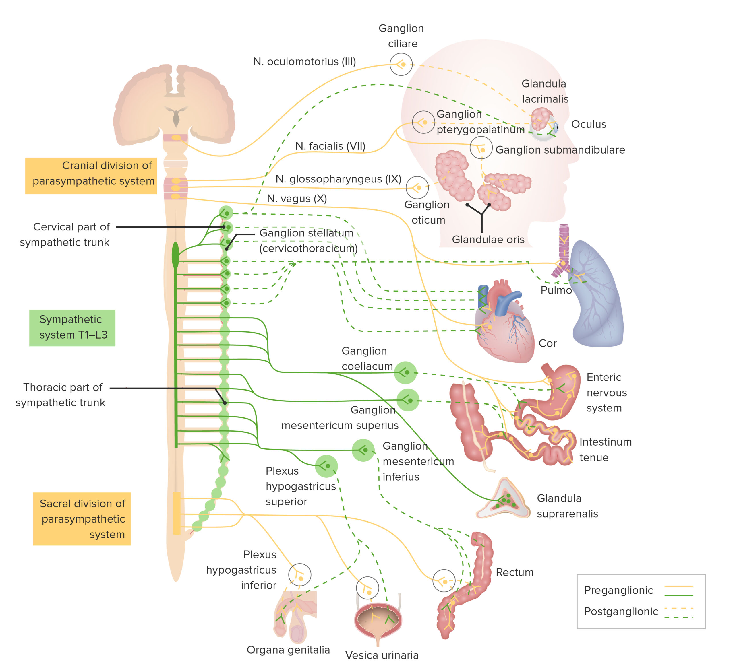
00:00 Well finally, let me just show you a picture of the wall of the gut. 00:04 You see on the left-hand side, on the very top dark pink area, that is part of the muscle wall of the gut. The material down below represents large folds in the gut and finally the gut mucosa. We will cover that in more detail when we talk about the gut in a later lecture. 00:25 But if you look very carefully in the middle of the wall of that muscle, the muscularis externa, you can see tiny little ganglion cells. They represent the enteric ganglia. That independent network of nerve cells that control the contraction of that musclaris externa muscle area as well as another muscle layer you can barely see. They are called the musclaris mucosa. 00:55 But again I will talk to you in detail about that when we look at the digestive system in a later lecture. 01:04 So in summary, it is important for you to be able to describe the motor pathways, both the somatic motorways and the visceral motor pathways. And also be able to recognize in the spinal cord what structural components those pathways relate to. Similarly you should know the sensory pathways, both the somatic sensory and the visceral sensory pathways. 01:34 And again also be able to locate within the spinal cord where those pathways travel and what structural components in the spinal cord relate to those sensory pathways. It is very important that you understand the functional significance of ganglia and also their structures. 02:01 And be able to differentiate between a sensory ganglion or dorsal root ganglion and ganglia belonging to the synthetic components of the autonomic nervous system and the parasympathetic components of the autonomic nervous system. So thank you for listening to this lecture.
About the Lecture
The lecture Enteric Ganglion by Geoffrey Meyer, PhD is from the course Nerve Tissue.
Included Quiz Questions
Where is the myenteric (Auerbach's) plexus of the gut wall located?
- Muscularis externa
- Epithelium
- Submucosa
- Muscularis interna
- Serosa
Author of lecture Enteric Ganglion

Geoffrey Meyer, PhD
Customer reviews
5,0 of 5 stars
| 5 Stars |
|
1 |
| 4 Stars |
|
0 |
| 3 Stars |
|
0 |
| 2 Stars |
|
0 |
| 1 Star |
|
0 |
It's An Good Overview And Histological Studies Of Ganglion.,But There is Need to Tell Stains Name
