Playlist
Show Playlist
Hide Playlist
Elbow Joint – Joints of Upper Limb

-
Slides 09 UpperLimbAnatomy Pickering.pdf
-
Download Lecture Overview
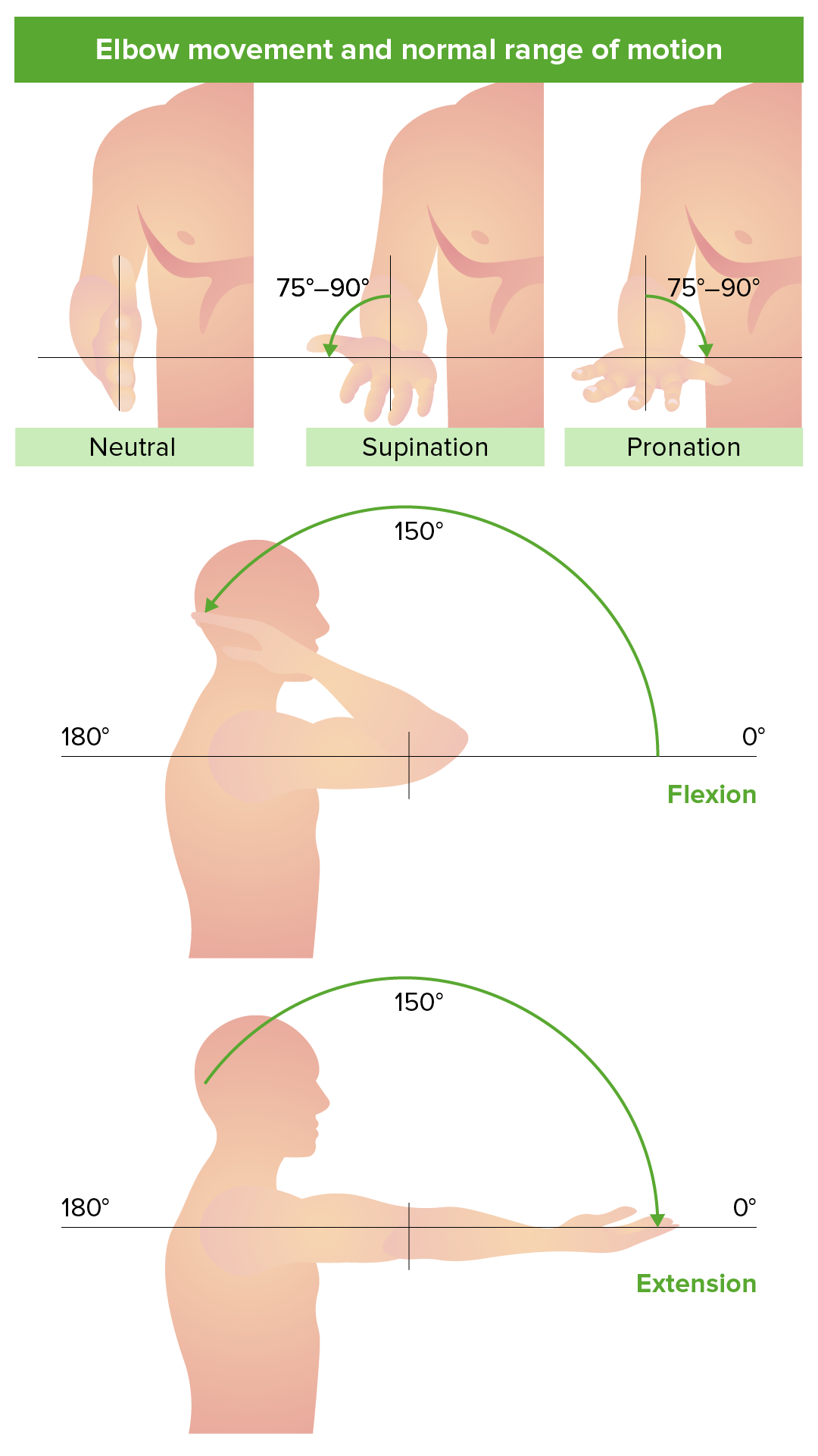
00:00 Now, let’s move on to the elbow joints and the articulation of the elbow joints. We have two articulations with the elbow joints, with the humerus articulating with both the radius and the ulnar. And here, we can see the anterior view of that joint, with the bones pulled apart to expose their articular surfaces. We can see we have the humerus here. We can see laterally, we have the capitulum of the humerus and that is going to articulate with the head of the radius. So the head of the radius is going to sit against the capitulum of the humerus. We then have the trochlea of the humerus which we can see here. And then remember, we have that C-shaped trochlea notch. We have the C-shaped trochlea notch that sits in this barrel-shaped trochlea. And these articulations allow flexion and extension to occur. The joint capsule around the joint surrounds the elbow joint and attaches to the edges of the articular surfaces. So it’s going to be running around these articular surfaces here. It can, in fact, cover both the coronoid fossa which we can see here. This is the coronoid process. And with full flexion, the coronoid process would sit in the coronoid fossa. The joint capsule will just go over there, so these articulations occur within the joint capsule, and it will also, as we’ll see, go over the olecranon fossa. And we can see that on this diagram here. Anteriorly, we can see the anterior view here. We can see the medial epicondyle and we see the lateral epicondyle. And we can see that the joint capsule is covering what would be the coronoid fossa here, with the coronoid process of the ulnar. And posteriorly, we can see we’ve got the olecranon fossa and the joint capsule is covering that olecranon fossa where it would house the olecranon of the ulnar. So we can see we’ve got the joint capsule running over the elbow joint. If we look at the ligaments that help to support the elbow joint, then we have a number of what are called collateral ligaments, and these are strong bands that reinforce the lateral aspect of the joints, that reinforce the lateral aspect of the joints. 02:29 And they are these lateral thickenings. We have the ulnar collateral ligament and we have the radial collateral ligament. The ulnar side we can see here, we’ve got the ulnar and we’ve got the medial epicondyle. And here we can see the ulnar collateral ligament. 02:49 These are triangular-shaped ligaments that are coming from the medial epicondyle of the humerus and passing to the coronoid process and they’re passing to the olecranon. 02:59 We can see them passing to the coronoid process anteriorly and then sweeping posteriorly, medial epicondyle. We can see them coming around onto the olecranon. These are your ulnar collateral ligaments, and these help to reinforce the joint. We have radial and these are fan like. We can see them on this radial side. Here’s the radius, and we’ve got this radial collateral ligaments. It’s coming from the lateral epicondyle, and it actually blends with a ligament known as the annular ligament, and that runs around the head of the radius. But these two collateral ligaments, these strong bands, that reinforce primarily the lateral aspect of the joint, the ulnar and the radial collateral ligaments. 03:46 The annular ligament, this encircles the head of the radius, and that is primarily involved in the proximal radio-ulnar joint which we’ll have a look at in a moment. But this joint allows flexion and extension to occur, and the joint capsule permits those movements between the capitulum and the radial head and the trochlea and the trochlear notch.
About the Lecture
The lecture Elbow Joint – Joints of Upper Limb by James Pickering, PhD is from the course Upper Limb Anatomy [Archive].
Included Quiz Questions
Which statement concerning the glenohumeral joint is correct?
- The coracoacromial arch prevents superior dislocation.
- The humeral head usually dislocates posteriorly.
- The coracohumeral ligament reinforces the joint inferiorly.
- The glenoid fossa articulates with more than half of the humeral head.
- The shoulder joint capsule is relatively tight and immobile.
Which statement concerning the glenohumeral joint is correct?
- The coracohumeral ligament strengthens the joints superiorly.
- The coracohumeral ligament strengthens the joints inferiorly.
- The coracohumeral ligament strengthens the joints anteriorly.
- The coracohumeral ligament strengthens the joints posteriorly.
Which statement concerning the elbow joint is correct?
- The radial/lateral collateral ligament is continuous with the annular ligament.
- The capitulum articulates with the notch of the trochlea.
- The coronoid fossa limits hyperextension.
- The carrying angle in women is approximately 25 degrees.
- Its bony components are the humerus and the radius.
Which bone is associated with the annular ligament?
- Radius
- Humerus
- Scaphoid
- Clavicle
- Scapula
With which movement does the coronoid process sit in the coronoid fossa?
- Flexion
- Extension
- Medial rotation
- Lateral rotation
- Supination
Author of lecture Elbow Joint – Joints of Upper Limb

James Pickering, PhD
Customer reviews
5,0 of 5 stars
| 5 Stars |
|
5 |
| 4 Stars |
|
0 |
| 3 Stars |
|
0 |
| 2 Stars |
|
0 |
| 1 Star |
|
0 |
