Playlist
Show Playlist
Hide Playlist
Echinococcus (Cestodes) – Helminths

-
03-60 Echinococcus.pdf
-
Download Lecture Overview
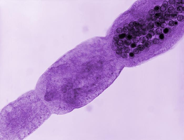
00:01 Echinococcus, parasites. 00:04 The Echinococcus come from the genus of Cestoda class, so they are helminthic like or worm-like. 00:11 In fact, they have a segmented body and appear more like a ribbon in nature. 00:16 Echinococcosis is classified as either cystic echinococcosis which is more common or alveolar echinococcosis. 00:24 Cystic echinococcosis or hydatid disease is caused by infection with the larval stage of echinococcus granulosus. 00:32 Most infections are asymptomatic. 00:34 It is common in sheep-raising areas of the Mediterranean, Middle East, Australia, New Zealand, South Africa and South America. 00:42 It is also found in both side of Canada, Alaska and California. 00:46 Dogs are the definitive hosts, having adult tapeworms in their gastrointestinal tract. 00:51 The intermediate hosts are herbivores, for example, sheep, horses and deer, as well as humans. 00:57 The tapeworm can cause the development of slowly enlarging, and potentially harmful cystic lesions in the liver, lungs and other organs. 01:05 Alveolar echinococcosis is caused by infection with the larval stage of Echinococcus multilocularis. 01:12 It is less common than cystic echinococcosis but it is a much more serious infection because the cysts are locally aggressive, causing destruction of surrounding tissue, and possibly leading to liver failure and death. 01:25 Adult E. multilocularis worms are present in foxes, coyotes, and dogs which are all definitive hosts. 01:33 The intermediate hosts are small rodents which harbor the hydatid larvae. 01:37 Infected dogs are the primary link to the occasional human infection. 01:41 This disease occurs mainly in Central Europe, Alaska, Canada, and Siberia. 01:47 In the United States, it is found in the Upper Midwest. 01:50 Other hydatid diseases are rare and cause cyst production primarily in the liver. 01:56 Two causative species are E. Vogelii and E. Oliganthus. 02:00 Both that which are found in Central and South America. 02:03 This slide shows the life cycle of echinococcus granulosus sensu lato or in the broad sense, since it includes several genotypes. 02:11 The adult echinococcus granulosus measures 2 to 7mm in length and resides in the small intestine of the definitive host, which is the dog. 02:20 The pregnant or gravid proglottids release eggs that are passed in the feces and which are immediately infectious. 02:27 After ingestion by a suitable, intermediate host, eggs hatch in the small intestine and release six-hooked oncospheres that penetrate the intestinal wall and migrate through the circulatory system into various organs, especially the liver and lungs. 02:42 In these organs, oncosphere develops into a thick walled hydatid cyst that enlarges gradually, producing protoscolices and daughter cysts that filled the cyst interior. 02:53 The definitive host becomes infected by injecting the cyst containing organs of the infected intermediate host. 03:00 After ingestion, the protoscolices evaginate, attach the intestinal mucosa and develop into adult stages in 32 to 80 days. 03:09 Humans are aberrant intermediate hosts and become infected by ingesting eggs. 03:14 Oncosphere are release in the intestine and hydatid cysts develop in the variety of organs. 03:20 If the cysts rupture, deliberated protoscolices may create secondary cysts in other sites within the body. 03:27 Known as "secondary echinococcosis." This slide shows the life cycle of Echinococcus multilocularis which causes alveolar echinococcosis. 03:35 The adult parasite measures 1.2 to 4.5mm in length and lives in the small intestine of the definitive host. 03:43 Gravid proglottids release eggs that are passed in the feces and they are immediately infectious. 03:48 After ingestion by a suitable intermediate host, eggs hatch in the small intestine and release a six-hooked oncosphere that penetrates the intestinal wall and migrates through the circulatory system into various organs primarily the liver as seen at E. Multilocularis. 04:04 The oncosphere develops into a multi-chambered thin-walled cyst that proliferates by successive outward budding which reflects it's locally aggressive character. 04:13 Numerous protoscolices develop within the cyst. 04:16 The definitive host becomes infected by ingesting the cyst containing organs of the infected intermediate host. 04:22 After ingestion, the protoscolices evagenates attach to the intestinal mucosa and develop into adult stages in 32 to 80 days. 04:31 Humans are aberrant intermediate hosts and become infected by ingesting eggs. 04:36 Oncospheres are release in the intestine and cysts develop within the liver. 04:41 Metastasis or dissemination to other organs, for example lungs, brain, heart or bone may occur if the protoscolices are released from cysts. 04:50 This sometimes is call "secondary echinococcosis." This next slide shows the case of a 40-year-old Syrian man with a 9 cm echinococcosis cyst due to E. Granulosus which was discovered incidentally during a chest CT examination. 05:05 The CT examination was essentially diagnostic because it showed multiple septae within the cyst and the patient was promen endemic region. 05:13 Suralgae was negative which is not unusual to see if the cyst is not leaked or ruptured which would expose echinococcus antigen the the immune system and create a response. 05:23 The patient underwent a left hepatectomy with the cyst removed intact. 05:27 The surgical specimen shows a multiloculated cyst filled with many daughter cysts and having a typically thick fibrous wall. 05:35 The microscopic photograph shows many protoscolices which account for the hydatid sand appears in the fluid which maybe seen on imagining. 05:44 The clinical disease caused by Echinococcus is cystic echinococcosis, and it is ingested or transmitted by ingestion of eggs in food contaminated with the eggs or food contaminated with dog or I suppose human feces. 06:02 The key is that the definitive host has to ingest the eggs. 06:07 Over then the period of months to years, the eggs might mature into their next step of larvae and create around them a cyst. 06:17 And this is not just any cyst, this is a large cyst with a very thin wall filled with clear fluid and it rarely or in fact almost never has any septa within it. 06:29 So it's a single compartment, large, fluid filled cyst. 06:33 Occasionally, a daughter cyst will bought off from the side and this even increases the size of this overall cyst. 06:41 Three forths, of the cysts occur in the liver and when so, they present with abdominal pain, certainly tenderness to palpation over the right upper quadrant. 06:50 The patient indeed may have a reactive hepatomegaly, liver size enlarging and they may even have what feels like a very firm mass along with that there may be some liver dysfunction causing jaundice and certainly there may be a low grade persistent fever. 07:09 In the lungs, the remainder of the cases almost one will have sort of non-specific respiratory symptoms so, a dry intermittent cough, a feeling of shortness of breath although rarely is there true failure of the respiratory status. 07:25 Patients may have referred chest pain as well. 07:28 Importantly, either of this locations whether it's in the liver the lungs or even in any place else in the body, has a huge potential for anaphylactic shock should those cysts rupture and in fact that is sometimes a clinical presentation when due to some other trauma, for example, a car accident and abdominal injury, the cyst rupture, delivers all of a sudden this huge flood of hypereosinophilic, anaphylactic type allergen and the body reacts in a heartbeat causing complete and total hyper IgE based reaction. 08:06 So, the treatment for this then is albendazole. 08:09 Again, a medication used primarily to treat helminthic or worm based disease. 08:15 Why not surgically remove this you ask? Because the risk of surgical - surgically induced rupture of these is just as severe as should that occur traumatically. 08:25 Sometimes if there is an actual, physical impediment, so, hepatomegaly or blockage of the common bile duct or blockage of the main set of bronchus, then surgery may be attempted but it is done in an incredibly careful and methodical fashion to remove the entire cyst together, with surrounding tissue and to avoid any, any risk, any even small risk of rupturing these. 08:50 So echinacoccus are very potentially dangerous if you come across them, perhaps the key things to remember here are sheep herders are high risk for those and they may present non-specifically and don’t, don’t, don’t, don’t, rupture those things. 09:07 This ends the lesson.
About the Lecture
The lecture Echinococcus (Cestodes) – Helminths by Sean Elliott, MD is from the course Parasites.
Included Quiz Questions
Which of the following organs is cystic echinococcosis most likely to involve?
- Liver
- Lungs
- Muscles
- Kidneys
- Brain
Which of the following species of Echinococcus is the most probable cause of cystic lesions in the liver?
- E. granulosus
- E. multilocularis
- E. vogeli
- E. oligarthrus
- E. equinus
Author of lecture Echinococcus (Cestodes) – Helminths

Sean Elliott, MD
Customer reviews
5,0 of 5 stars
| 5 Stars |
|
5 |
| 4 Stars |
|
0 |
| 3 Stars |
|
0 |
| 2 Stars |
|
0 |
| 1 Star |
|
0 |
