Playlist
Show Playlist
Hide Playlist
Development of the Midgut

-
Slides 07-45 Rotation and elongation of the midgut.pdf
-
Reference List Embryology.pdf
-
Download Lecture Overview
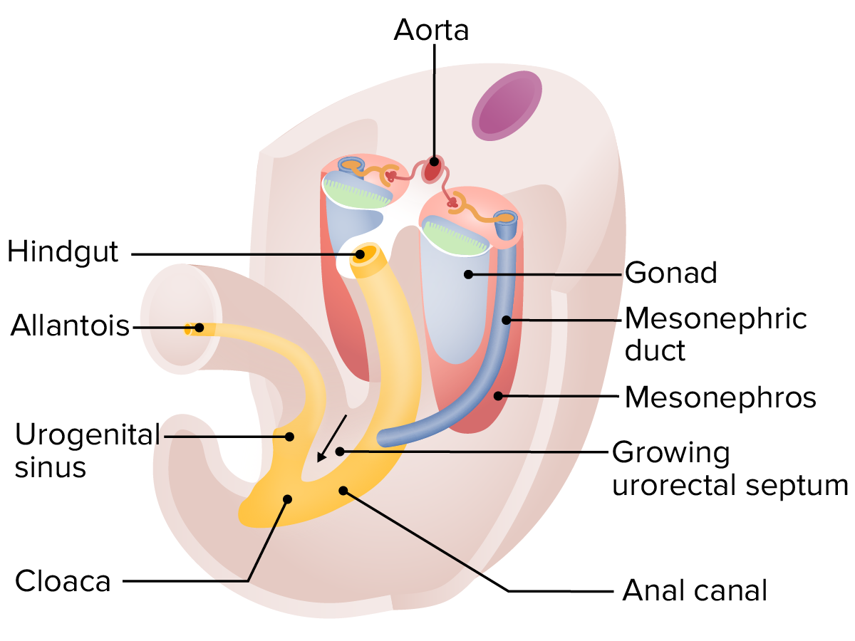
00:01 We will now discuss the development of the midgut and how it relates to the other portions of the gastrointestinal system. 00:07 The midgut begins as a U-shaped loop of the gut tube which stretches out along the umbilical cord and has the vitelline duct connecting it to the yolk sac, extending, continuing through the umbilical cord. 00:20 The midgut will become the distal portion of the duodenum, a portion of the pancreas, and then, the jejunum, ileum, cecum, vermiform appendix, the ascending and transverse colon and its blood supply comes through the superior mesenteric artery. 00:36 The major events that are going to occur in midgut formation are relatively simple in theory. 00:42 They are rotation and elongation. 00:45 The rotation of the midgut occurs along its blood supply. 00:49 The superior mesenteric artery acts as the axis for the rotation of the midgut and it undergoes a 270 degree rotation. 00:56 And if you were looking at me from the front as you are, that would appear to go in the counterclockwise direction. 01:02 The proximal cephalic loop, the part that’s on the top, is going to move inferiorly to become the distal duodenum, the jejunum, and the ileum. 01:11 The more caudal loop, the part on the underside is going to move superiorly into the right to become the rest of the small intestine, the ileum, as well as the cecum, appendix, ascending and transverse colon. 01:25 Now, in addition to rotating along that 270 degree axis, there’s gonna be a tremendous amount of elongation of the midgut at this point. 01:34 This elongation and just pure space that the midgut is taking up causes it to herniate out into the umbilical cord during week six. 01:43 During normal development however, the abdominal cavity will enlarge and the gut will return into the abdomen by week 10. 01:51 This is called a physiologic herniation and it is a normal event in development so long as it goes away. 01:58 If it maintains its presence in the umbilical cord for an extended period, that will be problematic. 02:04 Now, things that can go wrong is that you can have the gut rotate improperly and lay down in the wrong position. 02:11 Before we can understand the things that go wrong, let’s do a little more review on how the organs of the midgut wind up in their mature position. 02:20 So if we think about the midgut extending out from my umbilicus, I’m gonna take a hard right turn like I’m driving a car and the more distal portion in my left hand is going to move to the right side of my abdomen. 02:33 That’s why we find the cecum and appendix in the lower right quadrant of the abdomen. 02:38 Likewise, the more cephalic structures like the jejunum are going to be curling this way and lay down on the left side of my abdomen. 02:48 Typically, the cecum comes to rest in the lower right hand corner and the jejunum tends to rest in the upper left corner of my abdomen and the ileum is laid down progressively more to the right as time goes by. 03:00 As the cecum is moving into the lower right quadrant, it has the appendix bud off of it and elongate. 03:07 Because of that, the appendix is coming into existence as the cecum is coming into its final position. 03:14 And so, the appendix may be floating lose out in the mesentery, have its own attachment to the body wall, or it may be tucked under the cecum and relatively obscured. 03:25 That’s why finding an appendix when you’re trying to remove one can sometimes be problematic. 03:30 One thing to note is that there are smooth muscle bands on the surface of the large intestine called teniae coli and if you find them and follow them down to the base of the cecum, they always terminate at the vermiform appendix. 03:43 So if you’re having trouble finding it, locate one of the teniae coli and it’s your roadmap to the appendix. 03:49 As the gut tube reaches its final position and the midgut is going to get to the place where it belongs, portions of it will fuse to the posterior body wall and lose their mesentery. 04:02 The mesentery doesn’t literally disappear but as it folds back on the posterior body wall, it does fuse to a limited degree and those portions of the midgut are now immobile. 04:13 These are called secondarily retroperitoneal structures. 04:17 Secondarily because they didn’t used to be retroperitoneal but they became that way. 04:23 And this would be the ascending colon, the descending colon, as well as most of the duodenum, very little of the duodenum has a mesentery. 04:31 Structures that do have a mesentery like the jejunum, the ileum, the appendix, the sigmoid colon, and the transverse colon are all referred to as intraperitoneal structures. 04:42 Meaning, that they’re loose in the abdomen and suspended by a mesentery. 04:47 Now, just to fill out the end of that scheme, primary retroperitoneal structures are those that never existed in the abdominal cavity and were always located posterior to it such as the kidney and the adrenal glands and we’ll discuss those in their own separate lecture. 05:02 As the midgut is returning into the abdomen, it has the vitelline duct stretching to the yolk sac present coming off of the ileum. 05:10 Typically, that connection is going to disappear as the yolk sac disappears and there’ll be no trace of it. 05:17 However, you can have what’s known as an ileal or Meckel’s diverticulum. 05:22 In this case, there’s a tiny little pouch sticking off of the ileum which is a remnant of that vitelline duct and its connection to the yolk sac. 05:30 This is occurring in what we referred to as the rule of twos. 05:35 About 2% of the population are affected. 05:38 It affects males in a two to one ratio versus females. 05:41 You find it within two feet of the ileocecal valve where the ileum meets the cecum. 05:46 They’re usually approximately two inches long and you often find two types of mucosa in there. 05:52 Accessory gastric or pancreatic tissue can be found alongside normal intestinal tissue and generally, they’re going to occur and present before the patient is two years of age. 06:02 Now, these ileal diverticula can also be relatively clinically invisible. 06:08 They don’t always manifest and some people have them and don’t know it. 06:12 Problems that can occur are that occasionally, you don’t just have a little pouch. 06:16 You can actually have a little cord of tissue anchoring that ileal diverticulum to the umbilicus from within. 06:23 So it’s tethered to the anterior body wall. 06:25 The problem with this is that it can occasionally get twisted around that cord causing ischemia and volvulus. 06:33 Volvulus means twisting and in this case, it’s going to cause a great deal of pain as that portion of the gut is deprived of its blood supply and becomes ischemic. 06:42 Other problems that can occur is you can have little remnants of that connection if the vitelline duct maintain their connection to the ileum and form little cysts either at the umbilicus or just inside the body wall. 06:56 These may enlarge and become painful, and once again, serve as a locus for volvulus to occur if the intestine starts wrapping around that connection. 07:05 And you can also have what are referred to as vitelline fistulas. 07:09 A fistula is any inappropriate connection from one thing to another. 07:14 A connection that shouldn’t be there. 07:15 So a fistula is something that punched through from one space to another and in this case, an ileal diverticulum has a vitelline fistula connecting it to the umbilicus and you can guess how this is going to present. 07:28 The infant will have fecal material or partially digested food leaking out of his or her umbilicus. 07:35 Now, malrotation of the midgut is one other thing that can go wrong in the process of midgut development. 07:41 Remember, we typically have a 270 degree rotation like I’m turning my car to the right. 07:48 So counterclockwise from your view. 07:50 But sometimes, the gut only turns 90 degrees and then, moves back in. 07:55 In this case, the more distal portion of the midgut winds up on the left side. 08:00 So I’m going to have all of my large intestine and cecum packed into the left side of my abdomen and more proximal structures like the jejunum and ileum on the right side. 08:09 We can also have gut rotation go the opposite direction and go 270 degrees clockwise from your view or from my view. 08:18 I’m taking a hard left turn as I turn the steering wheel and my gut will wind up strangely in about the right place. 08:25 The organs will wind up more or less where we’d expect them to but the duodenum instead of being posterior to the transverse colon is going to be anterior to it and can sometimes block the transverse colon and cause some intestinal blockage and a bit of colonic obstruction. 08:41 Now, these malrotations are often asymptomatic and are found incidentally during abdominal operations or imaging. 08:48 Things that will manifest however when midgut rotations and malrotations go in very strange directions and wind up blocking the passage of food and fecal material through the intestines. 09:01 Mixed rotation where one segment rotates but the other does not can produce a variety of strange appearances to where the gut contents were laid down. 09:12 But the important thing to know is that all the gut components are still there, they’re just in the wrong place. 09:17 The problem is we can wind up with volvulus as one portion of the intestine wraps itself around another fixed structure, or another organ that’s in the wrong place. 09:27 This volvulus as we mentioned before with the ileal fistula and ileal diverticulum can wrap the intestine around itself, close off its blood supply and result in ischemia and death if it’s not treated. 09:41 Malrotation can also produce some problems where a portion of the gut that wants to be retroperitoneal attempts to fuse to the body wall and winds up blocking another portion. 09:53 If our gut rotates normally, the parts that are going to fuse to the posterior body wall have a relatively unobstructed pathway there. 10:00 If malrotation occurs, they may compress other portions of the gut on their pathway to seal themselves off with the body wall. 10:08 And as before, that can obstruct movement of fecal material and food through the gut or compromise the blood supply to the affected area. 10:16 Thank you very much for your attention and we’ll return with another topic.
About the Lecture
The lecture Development of the Midgut by Peter Ward, PhD is from the course Development of the Abdominopelvic Region. It contains the following chapters:
- Rotation and Elongation of the Midgut
- Vitelline Duct and Illeal Diverticulum
Included Quiz Questions
Which of the following does not originate from the midgut?
- Proximal duodenum
- Ileum
- Jejunum
- Cecum
- Ascending colon
Which of the following is true regarding the herniation and rotation of the midgut?
- The midgut rotates around the superior mesenteric artery.
- The midgut rotates around the inferior mesenteric artery.
- Due to the extensive elongation of the midgut, from weeks 10 to 16 it extends out of the abdomen into the umbilical cord.
- Herniation of the midgut into the umbilical cord is a pathologic process associated with polyhydramnios.
- The distal midgut loop rotates superiorly, forming the distal duodenum, jejunum, and part of the ileum.
Which of the following is false regarding the development of the appendix?
- The appendix begins to form once the cecum is in place in the right lower quadrant.
- The appendix may be located in several positions.
- The appendix begins to form as the cecum returns to the abdomen.
- The appendix forms between weeks 6 and 10.
- The location of the appendix may not have clinical relevance.
Which of the following statements regarding a Meckel’s diverticulum is false?
- They are typically 2 centimeters long.
- They affect about 2% of the population.
- There is a 2:1 male-to-female ratio.
- They are found within 2 feet proximal to the ileocecal valve.
- They usually present before 2 years of age.
Which of the following is false regarding malrotation of the midgut?
- If gut rotation occurs 270 degrees counterclockwise, the gut will appear completely normal except that the duodenum will run anterior to the transverse colon, which can lead to bowel obstructions.
- Any sort of malrotation increases risk of volvulus.
- Malrotation can result in adhesions.
- Malrotation can lead to compromised blood supply.
- Unusual organ location, like the cecum being fixed inferior to the stomach, may result from mixed rotation.
Author of lecture Development of the Midgut

Peter Ward, PhD
Customer reviews
4,0 of 5 stars
| 5 Stars |
|
0 |
| 4 Stars |
|
1 |
| 3 Stars |
|
0 |
| 2 Stars |
|
0 |
| 1 Star |
|
0 |
it is quite helpful ! i recommend it ! well explained and organized
