Playlist
Show Playlist
Hide Playlist
Cubital Fossa – Anatomy of the Arm

-
Slides 05 UpperLimbAnatomy Pickering.pdf
-
Download Lecture Overview
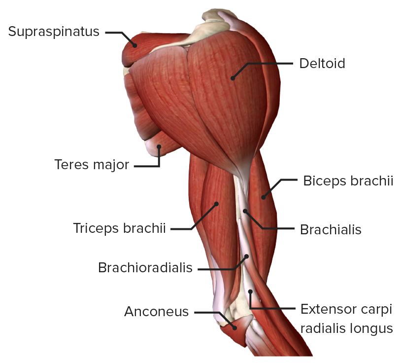
00:00 So if we look at the boundaries of the cubital fossa, these are made up of those muscular and bony boundaries. We've got superficial view here and we've got a slightly deeper view here. 00:13 This is the anterior aspect of the right arm, elbow and then we have got the forearm here. 00:21 We can see the muscle belly of biceps brachii. We can see brachialis. We can see running across the elbow joint we have got the bicipital aponeurosis and running down in this direction we will find those parts that run in the cubital fossa. If we just look at the boundaries to start off with the cubital fossa is the shallow triangular depression directly anterior to the elbow joint. 00:47 The boundaries superiorly is a horizontal line indicated here in green. Its really an imaginary line that is running between the two medial and lateral epicondyles. So it is running here. We then got the apex of the triangle being formed. Laterally by brachioradialis muscle we can see here and medially by pronator teres muscle we can see here. So if we continue these boundaries up to the superior boundary we can find a triangle and this is a cubital fossa. 01:21 The flow of the cubital fossa we can see in this deeper dissection we have brachialis muscle here and we also have another muscle known as supinator and this form the floor of the cubital fossa. So now brachioradialis here has been reflected, we can still make out pronator teres running in this direction. But because the bicipital aponeurosis has been reflected, we can see into the space of the cubital fossa. The contents of the cubital fossa: Well there is a nerve. There is an artery and there is a tendon. So here we can see on the right elbow joint the cubital fossa and from lateral to medial we find we have a tendon. We find we have an artery. We find we have a nerve. In a deeper dissection we can see the cut tendon, we can see the artery and we can see the nerve. So from lateral to medial we have tendon, artery and nerve. If we look the bicipital tendon we then have the brachial artery. 02:28 We then have the median nerve. We can see that in this deeper dissection the bicipital tendon has been cut. Bicipital tendon, brachial artery and median nerve. Running over the cubital fossa, we have the median cubital vein. Remember the median cubital vein was formed when the cephalic and the basilic veins were sending out for the upper limb and at the elbow there was this union between the two which is the median cubital vein. We can see the cut cephalic here and the basilic here and running across would have the median cubital vein. 03:07 We also have various cutaneous nerves following the basilic and the cephalic veins and these are supplying the forearm. So the contents to recap within the cubital fossa: We have the termination of the brachial artery, we can see here. If we follow the brachial artery deep through the cubital fossa we find we have it dividing into the radial and the ulnar arteries. With them we can see we have the median nerve. We can see we have the biceps tendon. If you were just to move brachioradialis to one side as reflected. We will see that we also have the radial nerve positioned here. Not really in the cubital fossa when its all intact, but by removing the brachioradialis we can expose the radial nerve. so the contents of the cubital fossa bearing in mind that for venupuncture the median cubital nerve is approached, then caution has to be taken by the medical practitioner that they don't pass the needle too far and start rupturing these structures.
About the Lecture
The lecture Cubital Fossa – Anatomy of the Arm by James Pickering, PhD is from the course Upper Limb Anatomy [Archive].
Included Quiz Questions
From medial to lateral, which structures are contained within the cubital fossa?
- Median nerve, brachial artery, and bicipital tendon
- Bicipital tendon, brachial artery, and median nerve
- Bicipital tendon, median nerve, and brachial artery
- Brachial artery, bicipital tendon, and median nerve
- Median nerve, bicipital tendon, and brachial artery
Which important vein runs over the cubital fossa?
- Median cubital vein
- Axillary vein
- Cephalic vein
- Basilic vein
- Ulnar vein
Which structure forms the medial boundary of the cubital fossa?
- Pronator teres muscle
- Brachioradialis muscle
- Median nerve
- Ulnar nerve
- Brachial artery
Author of lecture Cubital Fossa – Anatomy of the Arm

James Pickering, PhD
Customer reviews
5,0 of 5 stars
| 5 Stars |
|
1 |
| 4 Stars |
|
0 |
| 3 Stars |
|
0 |
| 2 Stars |
|
0 |
| 1 Star |
|
0 |
Good pictures Understanding the way he teaches Slow and steady
