Playlist
Show Playlist
Hide Playlist
Constrictive Pericarditis: Square Root Sign
-
Slides Pericardial Disease Constrictive Pericarditis Cardiovascular Pathology.pdf
-
Download Lecture Overview
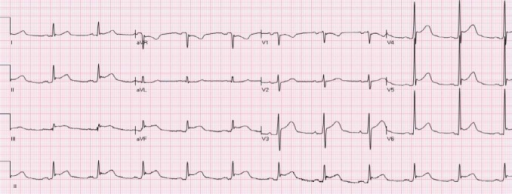
00:00 so that you’ll never missed it. Here we are. Let’s begin. What is this? Top curve. Tell me what that is? Left ventricle pressure curve? No. 00:10 Aortic pressure curve? No. Atrial tracing? Yes. How did you know? A and V curves. Why is this C not here? Why is this C-wave not here? 50 percent of your patients do not even have it. Clinically really. irrelevant, insignificant shall we say. Okay. So A and V wave. huge deals. X descent means downward. Y descent also means downward. Okay now. why are they down? Well, you tell me, A wave represents what? Late diastole. remember that our conversations we’ve had before. Late diastole last bit of blood kicking in and so therefore the atria is now empty. Decrease in pressure. Now. what should be super impost over that X descent and sufficiently moving in to the V wave? Should be the left ventricle pressure curve? Would you tell me. what part of our cardiac exact were in? Systole. Right? So the left ventricle was kicking up blood and the atria’s filling up with blood. If it the left atrium, pulmonary veins; right atrium obviously. IVC SCC. So this C is to always see this conversations that we’re having repetitively. So that we can just reinforces material. What then happens at V wave? Take a look at that S2. The S2 means closure of your aortic valve but then you move down isovolumetric relaxation and then the mitral valve should open and all that blood which is represented by the V-wave in your atrium is. whoop, gushing into the ventricle. Welcome to wide descent. Now, this is the problem. 01:56 In constrictive pericarditis, we have something to call this square root sign. in which there is so much blood that is now being rushed into the left ventricle. such a quick manner. Increasing pressure but the diastolic blood pressure is pretty much equalized throughout the chambers especially the ventricles. And that looks the square root sign because of constrictive pericarditis. And then you’ll notice immediately thereafter when the heart has been emptied or the atria’s filling has completely emptied into ventricles. That its pressure goes right back up. right backup. Welcome to constrictive, constrictive pericarditis. 02:33 In the meantime, take a look at the EKG down below and you’ll notice that atrial activity or the 02 quad activity and atria takes place first. For example, you see the P-wave there at the bottom. the P-wave there is the auto quad activity of the SA node kicking. Followed by the A wave which is the mechanic quad activity of the atria kicking. Is that clear? So as you move through here. you’ll see further for the EKG but the bottom-line is that’s square roots sign that you’re finding and which you find immediate increase in pressure due to your constrictive pericarditis called the square root sign. We’ll see one more time here. But in the form of. remember this. This suppose paradoxus. Would you tell me one more time. what should be the limit at what your systole blood pressure You should drop upon inspiration? 10, 10, 10, A drop in systole blood pressure upon inspiration of maximum of 10. If you go and you drop below 10. Or if you drop greater than 10 millimeters that this indicates your pulse paradoxus with inspiration. Is that clear? And these are conversations we had before increase amount of blood. And so therefore, you push your ventricle wall into left ventricle and that is what this is What you’re seeing here, pulse paradoxus. It was what with diffusion.
About the Lecture
The lecture Constrictive Pericarditis: Square Root Sign by Carlo Raj, MD is from the course Pericardial Disease: Basic Principles with Carlo Raj.
Included Quiz Questions
What does "a wave" represent in the venous waveform?
- Late diastole
- Early diastole
- Late systole
- Early systole
- Diastole and systole
What point in the venous waveform represents the closure of the aortic valve?
- v wave
- a wave
- x descent
- y descent
- c wave
Customer reviews
5,0 of 5 stars
| 5 Stars |
|
5 |
| 4 Stars |
|
0 |
| 3 Stars |
|
0 |
| 2 Stars |
|
0 |
| 1 Star |
|
0 |


