Playlist
Show Playlist
Hide Playlist
Congenital Melanocytic Nevi

-
Reference List Pathology.pdf
-
Slides Congenital Melanocytic Nevi Dermatopathology.pdf
-
Download Lecture Overview
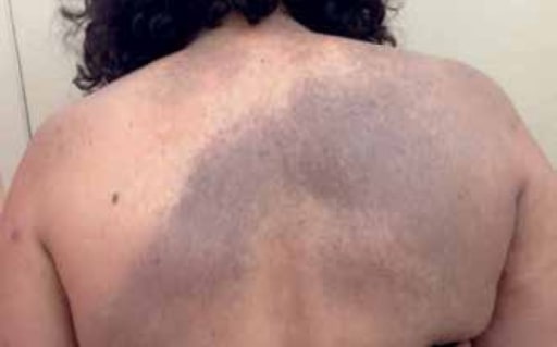
00:01 Welcome. Let's talk here about congenital melanocytic nevi or CMM. 00:07 And this is to be distinguished from acquired melanocytic nevi which will be the topic of a different talk. So cmms are just the common household mole. 00:18 They are pigmented skin lesions that are present at birth, hence congenital. And they're composed of a focal collection of proliferated melanocytes. 00:31 So the epidemiology of these they're actually quite common. 00:33 1 to 3% of newborns will have one maybe more. 00:38 Giant melanocytic nevi or congenital melanocytic nevi occur in about one inch 20,000. So the very big variety that we'll see at the end of the talk. 00:50 Kind of uncommon. But the regular household variant mole pretty common. 00:56 The pathophysiology of these. 00:58 So there is a genetic predisposition. 01:00 You can have somatic mutations in Braf which is a transcription factor. 01:06 It is a valine to glutamic acid mutation. 01:10 That's the V to E mutation in the 600th amino acid. 01:16 It's actually reasonably common in these. 01:19 You can also have in the large variety mutations in Nras, which is going to be a family that drives a family of genes that drive proliferation. 01:31 Notably, both Braf and Nras mutations are also going to be associated with melanoma. 01:38 I will say this now, but you should also keep in mind that these congenital versions almost never convert to into malignancy. 01:48 That is to say that driver mutations like Braf and Nras don't necessarily equate to malignancy. There are other changes that have to occur. 01:58 So what is this? What is this lesion. 02:01 It's just a proliferation of melanocytes and also migration of these proliferated melanocytes in deep to the basal layer and into the dermis. 02:13 The melanocytes of a congenital melanocytic nevus may migrate completely into the dermis, resulting in an intra dermal nevus as seen here on the right side. 02:26 The melanocytes start at the dermoepidermal junction and then gradually descend to become a junctional or compound nevus, present at both dermoepidermal junction and in the dermis, and then an intra dermal nevus as they mature. 02:42 Nivea have varying degrees of pigmentation and may be non-pigmented as seen here. 02:47 So for a clinical presentation, what do these look like? You as good doctors should be able to recognize them. 02:55 Most are small to medium sized and they tend to be solitary. 02:59 So when I say small 2 to 3mm up to maybe 1 to 2cm in size, depending on how much melanin is being produced by the cells, the color can range from tan to dark black. 03:12 They tend to have irregular borders, and because these occur in areas where hair would normally be Normally be produced, they may have hair within them. 03:21 So you have hairy nevi. 03:25 The large variant are often accompanied by multiple, smaller, widely disseminated kind of satellite nevi, and this is to be distinguished from similar acquired lesions. 03:35 So this is not the same as a Becker's nevus or Becker's melanocytic nevus. 03:41 And if you've never seen one, you can Google Richard Gere and look at his left shoulder. And there is a classic example of a Becker's that's to be distinguished, however, from these giant congenital melanocytic nevi, which are quite big with kind of satellite lesions. 03:58 How do we diagnose this? Well, it's kind of by inspection. 04:01 Um, you can also use a dermatoscope that will illuminate and give you a better sense of the kind of depth of invasion, etc., of, of the lesion. 04:12 And it can be very helpful in differentiating very small congenital melanocytic nevi from atypical acquired nevi. 04:20 I would leave the dermoscopic kind of evaluation to the cognoscenti, to people like dermatologists. 04:27 But you will undoubtedly use a dermatoscope in your clinical training. 04:30 And if you go into dermatology, you'll use it as part of your regular armamentarium. Uh, again, just to reiterate, the bulk of congenital melanocytic nevi are entirely benign. 04:45 They start benign and they end life benign. 04:48 The risk of melanoma arising in them is extremely low, despite the fact that many may be driven by proliferating specific mutations that cause increased proliferation. 05:01 How do we manage these? So it's mostly clinical observation. 05:05 Again very rarely they go malignant but the vast majority do not. 05:09 Um you may because of cosmetic reasons want to surgically excise them. 05:15 And they're certainly amenable to that. 05:17 You can also blast them with laser therapy. 05:20 So benign congenital melanocytic nevi are pretty common and pretty benign.
About the Lecture
The lecture Congenital Melanocytic Nevi by Richard Mitchell, MD, PhD is from the course Disorders of Maturation and Pigmentation of the Skin.
Included Quiz Questions
Which genetic mutation is commonly found in congenital melanocytic nevi but rarely leads to malignant transformation?
- BRAF, V600E mutations
- P53 deletion
- CDKN2A mutation
- KIT amplification
- PTEN deletion
Which feature best differentiates giant congenital melanocytic nevi from smaller variants?
- Presence of multiple satellite lesions
- Presence of hair within the nevus
- Irregular borders
- Dark black coloration
- Dermal invasion
Author of lecture Congenital Melanocytic Nevi

Richard Mitchell, MD, PhD
Customer reviews
5,0 of 5 stars
| 5 Stars |
|
5 |
| 4 Stars |
|
0 |
| 3 Stars |
|
0 |
| 2 Stars |
|
0 |
| 1 Star |
|
0 |
