Playlist
Show Playlist
Hide Playlist
Clinical Anatomy of the Glomerular Apparatus
-
Slides RenalClinicalAnatomy RenalPathology.pdf
-
Reference List Pathology.pdf
-
Download Lecture Overview
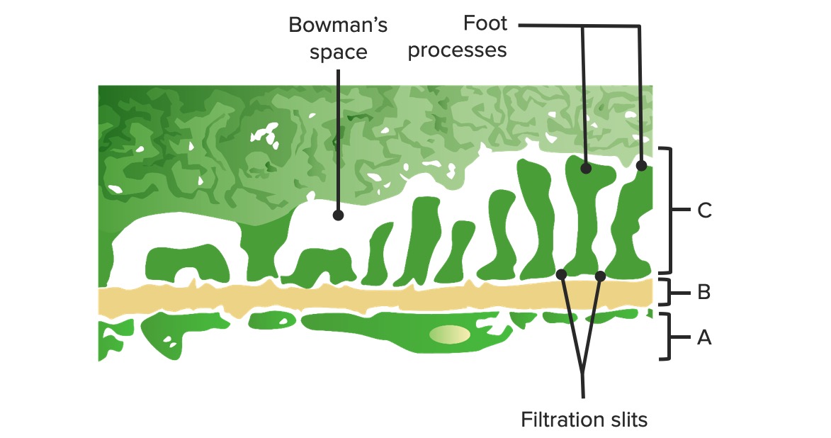
00:01 Let us talk further about our glomerular anatomy, that is relevant to us clinically. Endothelial cell. So what is that in the middle that we're seeing? You see two big circles. Those two big circles represent what? They represent glomerular capillaries. Next, what is the lining of the glomerular capillary? In other words, what is the lining of blood vessel? Always endothelial cell. What is beneath that endothelial cell? You see that dark blue line, the thickened blue line, that is your glomerular basement membrane. So in case you missed that picture, electron microscopy and you won't be able to make sense of it, maybe perhaps you must spend a little bit of time with this cartoon so that you clearly identifies where you are. By the way what type of imaging study could you see this in? Light microscopy, electron microscopy or immunofluorescence? This would be electron microscopy. So here, you find your glomerular basement membrane made up of what type of collagen? Good. Type IV collagen. What kind of charge? Negative charge. Things we have already talked about. That endothelial cells become really important to us because underneath that endothelial cell if you had a deposit, you will call it a subendothelial cell. Last little thing, we will take a look at as we progressed through here are going to be different components of this glomerular apparatus. Endothelial cell, may participate in the production of glomerular basement membrane. What you are seeing there, circle in green represents an endothelial cell. Initial segment of the filtration barrier, what does filtration mean to you? You are leaving those circles, who is? Plasma. In other words, really the water and the electrolytes and you are filtering through and then you're moving towards. You see this aliens? Looks like a one like a cyclops looks like an eye with feet on top of that circle. 01:57 You are moving towards that. What is that thing that we're seeing on top of that circle? Well that would be your visceral epithelial cell and a.k.a. you know that as being a podocyte. 02:08 Looks like a podocyte. It really looks like an alien though with a single eye. Imagine that thing crawling over your glomerular basement membrane. Let's identify more things. 02:17 Here is the glomerular basement membrane. Negative charge, collagen type IV. Glomerular basement membrane, microskeletons of the glomerulus. You give me a disease in which the type IV collagen is absent. Good. That is then called Alport, specifically type IV collagen deficiency. Be careful. As an important differential of collagen disease, you also have Ehlers-Danlos and you also have osteogenesis imperfecta, don't you? So those are three major collagen deficiency diseases that you definitely want to know before walking into your clinical wards or taking your boards. What were they again? Osteogenesis imperfecta, deficiency of collagen type I, Ehlers-Danlos, you have heard of plastic man right. And you have heard of hyperextensible joints. You have heard of skin being pulled out to all here and that is Ehlers Danlos and this is something in which we see this as being what? Output. 03:16 Glomerular basement membrane is important for you to know. Collagen type IV. Tram track we will talk about that as well. It participates obviously in filtration barrier. There it is. There is alien that I was referring to and that your visceral epithelial cell, give me its proper name that you want to know. A podocyte. Now, what are those little feet underneath that podocyte, underneath that little blob? Those are your foot processes. 03:41 In between the foot processes, what would you have? Slit diaphragms so that you have proper filtration of your plasma from the blood vessel, through your endothelial cell fenestrations, through the basement membrane and to the side of the Bowman space. Visceral epithelial cell. What if you find deposit underneath that epithelial cell? You call that a subepithelial deposit. 04:05 Prototype, PSGN. Produces glomerular basement membrane, intercellular junctions are the final filtration barrier. Once you get passed this, you are now officially ladies and gentleman in the Bowman space. One little cell that I want to make sure that you are clear about, you see the lining on top there. You see the lining on top all way outside of your cells. That is your parietal epithelial cell. And so therefore if these are proliferating in a condition are called RPGN, rapidly progressive glomerular nephritis, you will call that your crescentic cells. In the middle there is a mesangium, what does that mean? Think of this as being your smooth muscle. Why do I keep saying that? Because at some point in time, if you glomerular are active, they are going to contract. Amazing, these things are and in the mesangium, it may then as we said become thickened and as you do so, you have deposition of your immune complexes such as IgA. Give me the two major differentials that we discussed. Good. IgA vasculopathy, a.k.a. Henoch-Schonlein purpura and the other one was IgA nephropathy a.k.a. Berger. Contraction, produces a matrix, responsible for well, protecting the entire area. Phagocytic. Mesangial matrix as what you see deposition of various things could occur in there. It is the supporting framework of the structures. The fenestra represents what? The holes between the endothelial cell, so you can have filtration taking places.
About the Lecture
The lecture Clinical Anatomy of the Glomerular Apparatus by Carlo Raj, MD is from the course Renal Clinical Anatomy.
Included Quiz Questions
What is primarily responsible for the negative charge on the GBM which repels albumin from being filtered?
- Heparan sulfate
- Podocytes
- Cationic proteins
- Inflammatory mediators
- Type IV collagen
What substance is increased in the development of Kimmelstein-Wilson nodules?
- Type IV collagen
- Heparin sulfate
- Immune complexes
- Albumin
- Type II collagen
Proliferation of ________ is responsible for the crescent formation in rapidly progressive glomerulonephritis?
- Parietal epithelial cells
- Type IV collagen
- Visceral epithelial cells
- Glomerular basement membrane
- Mesangial cells
Which of the following statements about subepithelial deposits is INCORRECT?
- Immune complexes that are deposited are produced in mesangial cells.
- It is a cause of glomerular basement membrane thickening.
- All the other responses are correct.
- Post-streptococcal glomerulonephritis is a potential etiology.
- They are associated with membranous glomerulopathy.
Which of the following is NOT a function of visceral epithelial cells?
- Supporting glomerular capillaries
- Producing the glomerular basement membrane
- None of the functions listed here is correct.
- Serving as a distal barrier to prevent protein loss in the urine
Which of the following cells is responsible for production of the glomerular basement membrane?
- Visceral epithelial cells
- Mesangial cells
- Fibroblasts
- Fenestrated cells
- Parietal epithelial cells
Which of the following diseases involves defective type IV collagen production?
- Alport disease
- Ehlers-Danlos syndrome
- All have defects of type IV collagen.
- Osteogenesis imperfecta
- Marfans
Which of the following substances is NOT able to permeate the glomerular basement membrane under physiological conditions?
- Strongly negatively charged proteins
- Water
- Proteins < 70,000 daltons
- Sodium
- Urea
Which of the following statements about mesangial cells is INCORRECT?
- They are the lining cells of Bowman's capsule.
- They are contractile.
- They are the site of immune complex deposition in IgA glomerulopathies.
- They are phagocytic.
- They produce the mesangial matrix.
Customer reviews
5,0 of 5 stars
| 5 Stars |
|
5 |
| 4 Stars |
|
0 |
| 3 Stars |
|
0 |
| 2 Stars |
|
0 |
| 1 Star |
|
0 |


