Playlist
Show Playlist
Hide Playlist
Case: 56-year-old Man with Headache

-
Slides CNS Infections Cerebritis.pdf
-
Download Lecture Overview
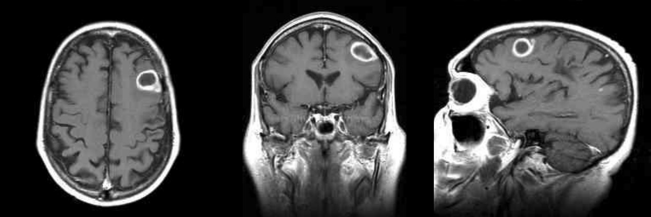
00:00 In this lecture, we're going to learn about cerebritis. Let's start with a case. A 56-year-old man with headache. This is a 56-year-old man with diabetes mellitus who does not follow with any physician and presents with new headache and aphasia, problems with getting the words out. This began about 1 week ago and he has worsened steadily. He has had intermittent fever, severe tooth pain, and there is concern for a periapical abscess in and around the first right maxillary molar. Examination shows an expressive type aphasia, he can't get his words out, with impaired fluency, intact comprehension to commands, and intact repetition. There is frontal lobe dysfunction. The temporal lobe appears normal and this looks like a transcortical motor dysfunction with intact repetition and naming. Otherwise, neurologic exam is unremarkable. MRI shows an enhancing lesion in the left frontal lobe with surrounding edema. So what's the diagnosis? Well let's walk through and dissect to this case. This patient needs imaging. There is some process that's been going on that sounds like it's infection. 01:12 There is a periapical abscess, a potential nidus in the maxilla to infection in the brain. 01:19 Examination shows a focal examination, a focal neurologic deficit with this expressive type aphasia, and that requires imaging. And the imaging shows an enhancing lesion in the left frontal lobe which we'll take a look at in the next slide. So here is this patient's imaging. 01:38 We're looking at a T2 weighted image, a T1 weighted image with gadolinium contrast, and then on the far right we see the diffusion weighted image. So what do you see? On the left, I see a lesion in the brain with surrounding vasogenic edema. In the middle, we see the periphery of this lesion is enhancing with contrast and we know that things that enhance with contrast are acute, infections, inflammation, and malignancy. This patient's clinical presentation is not consistent with malignancy which would be lower on our differential. Inflammation or MS would be uncommon to present in this way. And infection is at the top of our differential. 02:19 The diffusion weighted image shows prominent diffusion restriction in the center of this lesion, that necrotic core that you see on the gadolinium image and that suggest infection and points us strongly towards an abscess. So what's the diagnosis? Well, this doesn't look like meningitis, encephalitis, or cerebritis. The imaging is most consistent with brain abscess and this is a typical presentation, a patient with mild low grade fevers, a new focal neurologic deficit, and a potential nidus for seeding infection into the brain.
About the Lecture
The lecture Case: 56-year-old Man with Headache by Roy Strowd, MD is from the course CNS Infections.
Included Quiz Questions
Which of the following is suggestive of brain abscess?
- Fever, headache, and focal neurologic deficit
- Fever, headache, and diffuse cortical dysfunction
- Fever, headache, and meningismus
- Fever, headache, and altered mental status
- Headache, malaise, and myalgia
Which of the following MRI findings is suggestive of cerebral abscess?
- Ring-enhancing lesion with surrounding edema
- Multiple ring-enhancing lesions
- Periventricular calcification
- Basal ganglia calcification
- Combination of hypo-/hyperintense foci in the cerebral white matter
Author of lecture Case: 56-year-old Man with Headache

Roy Strowd, MD
Customer reviews
5,0 of 5 stars
| 5 Stars |
|
5 |
| 4 Stars |
|
0 |
| 3 Stars |
|
0 |
| 2 Stars |
|
0 |
| 1 Star |
|
0 |
