Playlist
Show Playlist
Hide Playlist
Axillary Artery – Axilla and Brachial Plexus

-
Slides 04 UpperLimbAnatomy Pickering.pdf
-
Download Lecture Overview
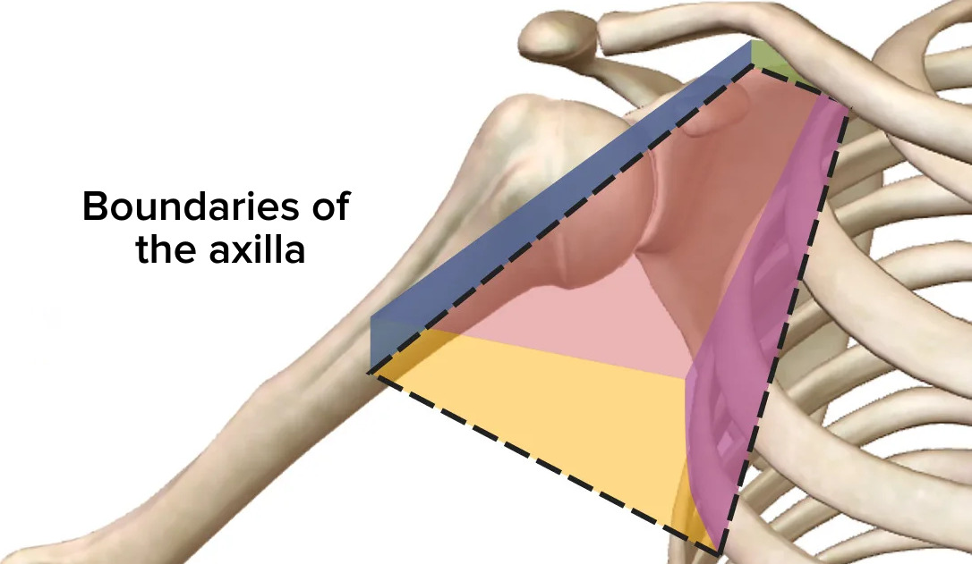
00:01 If we look at the contents of the axilla, then the main blood vessel is the axillary artery. And the axillary artery is a direct continuation of the subclavian artery as it passes into the axilla. Here now, we can clearly see the cervico-axillary canal. We can see anteriorly here, we have the clavicle. We can see medially, we have the first rib. And then posteriorly running along the back here, we have the superior border of the scapula. So here is the posterior boundary of the cervico-axillary canal. Here’s the anterior boundary and here is the medial boundary. And this forms the apex of the axilla which allows the axillary artery to pass in. 00:51 The axillary artery is a direct continuation of the subclavian artery as it passes into the axilla. Now, we can recognize three parts to our axillary artery. And these are associated with pectoralis minor muscle. Here, we have pectoralis minor muscle coming from the ribs and passing to the coracoid process of the scapula. The first part of the axillary artery is between the clavicle and this superior border of pectoralis minor. The second part is directly deep to pectoralis minor. And the third part is from this inferior boundary of pectoralis minor all the way down to the lower border of the axilla where the axillary artery then becomes the brachial artery. And that’s typically the inferior border of teres major. So if we have a look at the three parts of the axillary artery, then as I said, it’s a direct continuation of the subclavian artery starting at the lateral border of the first rib, and it ends at the inferior border of teres major. Divided into three parts. And each part gives rise to numerous blood vessels. So coming from the first part, there’s going to be one. Coming from the second part, there’s going to be two. And coming from the third part, there’s going to be three blood vessels. 02:20 So, if we look at the first part of the axillary artery, this is between the clavicle and the superior border of pectoralis minor. We have one blood vessel. And we can see that here. 02:35 That is the superior thoracic artery. Superior thoracic artery, going to supply the superior aspect of the thoracic chest wall. If we look at the second part, which is posterior to pectoralis minor, we have two blood vessels. We have the thoraco-acromial artery and the lateral thoracic. We have the thoraco-acromial artery which we can see here, and we also have the lateral thoracic artery which we can see here. And these are the two branches that come from the second part of the axillary artery. The thoraco-acromial artery is a small little root that then gives rise to a whole series of blood vessels, the acromial branch, the deltoid branch, various branches that go to supply this region. And we’ll detail this more when we look at the whole blood supply to the upper limb. Within the axilla, we then have the third part of the axillary artery. Within the axilla, we then have the third part of the axillary artery, and this gives rise to three blood vessels, the subscapular artery, the anterior and posterior circumflex humeral arteries. So the subscapular artery, we can see the subscapular artery running down in this direction, and we can see that may well go on to form various anastomosis around the scapula. We can see it running up in this direction. So anastomos here with a branch that’s coming off the thoraco-acromial artery. So a complicated anastomosis is going on, and like I said, we’ll come back to it. 04:15 That’s one of the branches coming from the third part of the axillary artery. 04:21 The remaining two branches to come off the third part are the anterior and posterior circumflex humeral arteries. Remember the posterior circumflex passes out through the quadrangular space. 04:32 It is accompanied by the axillary nerve. And these form an anastomosis around the surgical neck of the humerus. So they are the various branches of the axillary artery. Three parts, one artery coming from the first part, two arteries coming from the second, the three arteries are coming from the third. So an easy way to remember those arteries. 04:57 If we look at the lymph nodes, then there’s a whole series of lymph nodes that are located within the axilla. I have a whole series of axillary lymph nodes. And these axillary lymph nodes are going to receive the lymphatic vessels from other neighbouring lymph nodes. So for example, there are five groups of lymph nodes that drain into the axillary lymph nodes. We have pectoral lymph nodes from the anterior thoracic wall. We have subscapular lymph nodes from the posterior thoracic wall and scapula. We also have humeral lymph nodes from most of the upper limb. So, most of the limbs from the upper limb are draining into these humeral lymph nodes. We have central lymph nodes, and these are beginning to receive the various lymphatic vessels from the pectoral subscapular and humeral. So those three giving rise to lymphatic vessels that go to this central lymph nodes. And then from the central lymph nodes in the axilla, they are going to pass up to the apex of the axilla into apical lymph nodes. So we have these groups within the axilla that are receiving lymphatic vessels from the neighbouring regions. So pectoral subscapular and humeral, they pass to these central lymph nodes, and the central lymph nodes then pass up into the apex of the axilla. 06:25 This will then drain into subclavian lymph nodes, and ultimately, into the venous system. 06:30 So a very basic overview there. I don’t really want to get broke down in too much of the detail. But it’s important that from this whole kind of chest in upper limb region, they all pass to one central lymph node which then passes through the axilla to the subclavian lymph node, and ultimately, draining into the venous system.
About the Lecture
The lecture Axillary Artery – Axilla and Brachial Plexus by James Pickering, PhD is from the course Upper Limb Anatomy [Archive].
Included Quiz Questions
Which muscle is associated with the division of the axillary artery into 3 parts?
- Pectoralis minor
- Pectoralis major
- Biceps brachii
- Triceps brachii
- Serratus anterior
The second part of the axillary artery gives off which of the following?
- Lateral thoracic artery
- Superior thoracic artery
- Subscapular artery
- Anterior circumflex humeral artery
- Posterior circumflex humeral artery
Lymph from the posterior thoracic wall and scapula drain into which of the following?
- Subscapular lymph nodes
- Pectoral lymph nodes
- Humeral lymph nodes
- Central lymph nodes
- Apical lymph nodes
Author of lecture Axillary Artery – Axilla and Brachial Plexus

James Pickering, PhD
Customer reviews
5,0 of 5 stars
| 5 Stars |
|
5 |
| 4 Stars |
|
0 |
| 3 Stars |
|
0 |
| 2 Stars |
|
0 |
| 1 Star |
|
0 |
