Playlist
Show Playlist
Hide Playlist
Axilla – Axilla and Brachial Plexus

-
Slides 04 UpperLimbAnatomy Pickering.pdf
-
Download Lecture Overview
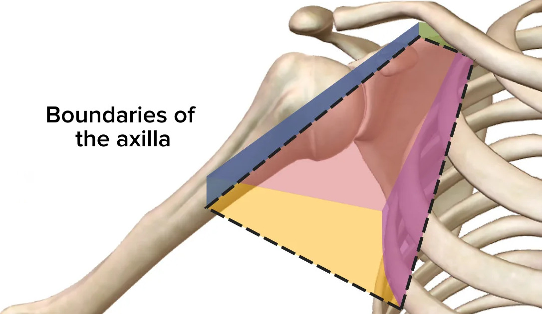
00:01 In this lecture, we're going to look at the axilla and the brachial plexus. 00:05 For the axilla which is a space between the humerus and the thoracic wall. 00:12 We're going to look at its boundaries. 00:14 We'll look at the walls, we'll look at its base, we'll look at the apex of the axilla. 00:19 We'll then look at the contents, the axillary artery, the axillary vein and some axillary lymph nodes and we'll also may attention to the brachial plexus which is within the axilla. 00:30 We'll then look at the brachial plexus itself. 00:32 We'll look at the various parts, its components and we'll look at the anatomical relation. 00:37 The axilla is a space that's located between the humerus and the chest wall. 00:45 Here we can see the humerus and we can see in this space here between the humerus laterally and the chest wall medially we find the axilla. 00:56 It's a pyramidal space and most of its boundaries are formed by muscles. 01:01 The axilla would assume this space in here medially we have the chest wall and serratus anterior and laterally in this abducted arm, we'll see that the lateral boundary is the intertubercular groove of the humerus. 01:19 We're looking in this space here, and as I mentioned, it's got a lateral and medial and anterior or posterior boundary for these walls. 01:29 It's got an apex and it has a space. 01:32 A pyramidal space that is below the glenohumeral joint between the humerus and the chest wall. 01:39 It provides an important passage way for vessels and nerves to pass to and from the upper limb, structures like the axillary artery, axillary vein and like I mentioned the brachial plexus. 01:52 The apex of the axilla is known as the cervico-axillary canal and this is located between the neck and the axilla. 02:02 The neck being between the head and your trunk and your axilla, cervico-axillary canal. 02:08 This canal is bounded by rib 1, the clavicle and the superior border of the scapula. 02:16 The clavicle, rib 1 and the scapula formed the boundaries of this apex. 02:22 The anterior wall is formed by both pectoralis major and minor as they're running from the chest wall to the humerus and the scapula and the posterior wall is formed by subscapularis muscle, the scapula, bone, teres major and latissimus dorsi muscle. 02:43 The lateral wall is formed by the intertubercular groove of the humerus and the medial wall is formed by the thoracic wall and serratus anterior muscles that lie on its lateral aspect. 02:55 The base is the concave skin joining the upper limb to the trunk. 03:01 Those axillary folds of skin that pass from the trunk medially to the arm laterally. 03:10 You can physically hold on to these pieces of skin as they're passing from the chest wall to the upper limb. 03:18 This formed the base and you can put your hand therefore within this folds of skin and that is your armpit. 03:27 We can see this in a bit more detail here, so here we have the cervico-axillary canal, we've got the clavicle here. 03:35 Posterior to the clavicle, we'd find the cervico-axillary canal and that contains those structures that are passing from the neck into the axilla. 03:45 We'd see rib 1 running around here and we've got the clavicle and then most posteriorly we've had the superior boarder of the scapula. 03:53 Anteriorly, we can find we have pectoralis minor, muscle here and here is the cottage of pectoralis major muscle and these are passing across towards even the humerus or the scapula. 04:06 Here, they are forming this anterior boundary. 04:09 Posteriorly, we have these muscles here, we have subscapularis, we can also see we have teres major running down here and we can see latissimus dorsai. 04:20 Posteriorly, we have subscapularis, we have teres major and then we have latissimus dorsai. 04:27 Laterally, we can see have the intertubecular groove that's running along the humerus with the various muscles inserting into the intertubercular groove. 04:39 And then medially, we see we've got this digits of serratus anterior, forming the medial boundary. 04:46 Anteriorly, we've got our pectoralis muscles, posteriorly, we've got subscapularis, teres major and latissimus dorsai. 04:57 Laterally, we've got the intertubercular groove and medially, we've got serratus anterior. 05:03 Structures going to pass in to the axilla via the cervico-axillary canal. 05:09 Within the posterior wall of the axilla, there are series of spaces and these are important spaces as they allow structures to leave the axilla and pass to the various locations so they can pass to the scapula region, they can pass to the posterior aspects of the arm like it can pass to the shoulder joint. 05:32 Within the axilla along its posterior wall, we have three spaces, these known as the quadrangular space, the triangular space and the triangular interval. 05:42 And these spaces are formed by the muscles and sometimes the humerus that forms spaces that allows structures to pass through. 05:51 The origin of these structures comes from deep within the axilla which has the main content being your axillary artery and the axillary vein. 06:00 Blood vessels from the axilla can pass out to neighboring structures through the spaces. 06:07 It also contains some axillary lymph nodes and it contains the brachial plexus. 06:12 Nerves can also pass out through these spaces.
About the Lecture
The lecture Axilla – Axilla and Brachial Plexus by James Pickering, PhD is from the course Upper Limb Anatomy [Archive].
Included Quiz Questions
Which statements about the boundaries of the axilla are correct? Select all that apply.
- The apex is formed by the clavicle, 1st rib, and superior border of the scapula.
- The medial wall is formed by the pectoralis major muscle.
- The anterior boundary is formed by the pectoralis major and minor.
- The posterior wall is formed by the subscapularis muscle, the scapula bone, teres major, and latissimus dorsi muscle.
- The lateral wall is formed by the intertubercular groove of the humerus.
How many important spaces are present within the axilla along the posterior wall?
- 3
- 2
- 4
- 5
- 6
Which structures are included in the contents of the axilla? Select all that apply.
- Axillary artery
- Pectoralis major and minor muscles
- Axillary vein
- Brachial plexus
- Axillary lymph nodes
Author of lecture Axilla – Axilla and Brachial Plexus

James Pickering, PhD
Customer reviews
5,0 of 5 stars
| 5 Stars |
|
1 |
| 4 Stars |
|
0 |
| 3 Stars |
|
0 |
| 2 Stars |
|
0 |
| 1 Star |
|
0 |
Best lecture ever, I never thought I'd be able to understand brachial plexus
