Playlist
Show Playlist
Hide Playlist
Autonomic Ganglion
-
Slides 11 Types of Tissues Meyer.pdf
-
Reference List Histology.pdf
-
Download Lecture Overview
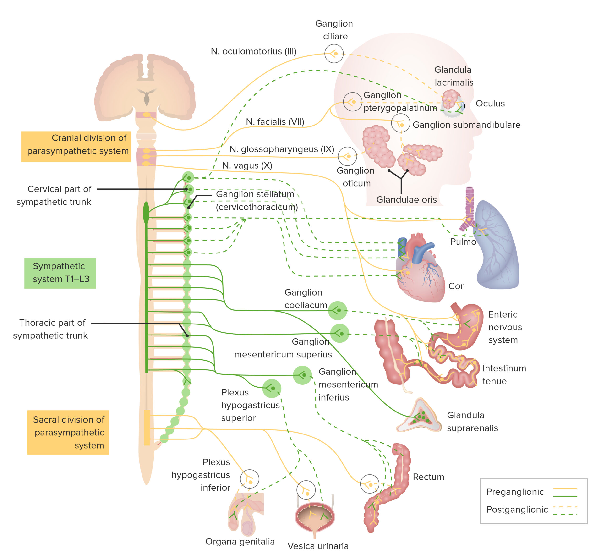
00:01 Well, on this diagram here, it is a picture or an image of one of these postsynaptic neurons in a paravertebral ganglia or part of the sympathetic ganglia. And there, you don't see a lot of satellite cells around the cell bodies. And that is because as I explained before, these neurons have a dendritic tree, communicating with preganglionic neurons. And because of that vast dendritic tree, the satellite cells cannot pack around as much as they do in a sensory or a dorsal root ganglion. Here is an example of a parasympathetic autonomic ganglion. Remember I said they are located out near the viscera, so that the postganglionic neuron travels a very short distance to the target tissue. And they are very hard to see sometimes. They are very tiny. This is a section in the tongue and you see a neuron rather a large structure surrounded by a few satellite cells embedded in the connective tissue areas of the tongue. Those postsynaptic neurons, the cell bodies you see labelled there, again they travel a short distance and innervate glands in the tongue shown here. Serous secreting glands that secrete materials that flush out fluid away from our taste buds so that we can continually taste different tastes. It is all controlled by secretion of the serous glands or controlled by the short postganglionic neurons travelling from postganglionic areas out in the periphery.
About the Lecture
The lecture Autonomic Ganglion by Geoffrey Meyer, PhD is from the course Nerve Tissue.
Included Quiz Questions
What type of autonomic ganglions are often found within the target organs?
- Parasympathetic
- Sympathetic
- This primarily depends on the type of organ.
- Autonomic ganglia are not found in peripheral organs.
Customer reviews
5,0 of 5 stars
| 5 Stars |
|
5 |
| 4 Stars |
|
0 |
| 3 Stars |
|
0 |
| 2 Stars |
|
0 |
| 1 Star |
|
0 |


