Playlist
Show Playlist
Hide Playlist
Asbestos Exposure and Pleural Disease

-
Slides 09 PleuralDiseases RespiratoryAdvanced.pdf
-
Download Lecture Overview
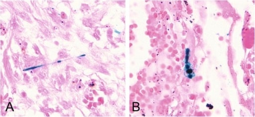
00:00 Right. Just a brief now, we discussed asbestos exposure in pleural disease because another cause of the pleural thickening is asbestos exposure. Occupational exposure asbestos used to become in the ‘70s and the early ‘80s mainly in the building trades but also engineers, Dockers and shipbuilders as asbestos was used as a heat prevention or heat protection substance in those industries. Inhaled asbestos fibres actually readily reached the alveoli, penetrate through the alveoli, and reached the pleural lining where they cause inflammation. And the consequences of that are three or four-fold. 00:38 One is that you get pleural plaques and this show off on a chest X-ray frequently in patients who’d been exposed to asbestos. In fact, really don’t mean much for the health but do identify patients who’ve had pleural who have been exposed to asbestos. 00:52 Benign pleural thickening where you get a layer of thickening around the lungs and that can cause restrictive lung defects. As I mentioned earlier, you can get benign exudated pleural effusions due to asbestos exposure. And the last thing that pleural asbestos can cause is a malignant mesothelioma which I discussed in the lecture of malignancy. Asbestos inhalation also can cause pulmonary fibrosis, and that’s called asbestosis, and that’s discussed in the lecture on Interstitial Lung Diseases. So to summarize the main learning points of pleural disease; pleural effusions are common and are divided into exudates in which the pleural is abnormal and transudates in which the pleura is normal, but there are other systematic reasons why pleural fluid is forming such as cardiac failure. Investigation of pleural fluid is best done using ultrasound as they identify exactly where the fluid is. 01:43 You can see loculations, and you see whether there are any abnormalities of pleural suggestive of cancer, etc. There are multiple different causes of pleural effusions, but the most important are malignancy either secondary metastasis or mesothelioma or pleural infection, TB or bacterial infection. If somebody has recurrent effusions and that occurs frequently in patients with metastatic disease affecting the pleura, then these can be prevented by pleurodesis where you fuse the visceral and the parietal pleura together. 02:13 If somebody has loculated pleural fluid, that suggests strongly that they have pleural infection and requires active treatment and investigation. Pneumothoraces, air in the pleural space could be primary young people with no underlying lung disease, but spontaneous holes appearing in the visceral pleural allowing air out into the pleural space or secondary where they actually have an underlying lung disease such as COPD or cystic fibrosis. 02:39 Pneumothorax can be relatively easily treated in most cases, but the serious problem is the tension pneumothorax which is a medical emergency with a high pressure in the pleural spaces causing hypertension and can lead to cardiac arrest. That needs immediate treatment by insertion of wide bore canula into the second intercostal space, midclavicular line which equalizes the high pressure in the pleural space without atmospheric. And thank you for listening.
About the Lecture
The lecture Asbestos Exposure and Pleural Disease by Jeremy Brown, PhD, MRCP(UK), MBBS is from the course Pleural Disease.
Included Quiz Questions
Which of the following is NOT a characteristic CT finding in asbestosis?
- Consolidation
- Pleural thickening
- Pleural plaque
- Pleural fibrosis
Which lung condition is a medical emergency?
- Tension pneumothorax
- Spontaneous pneumothorax
- Mesothelioma
- Emphysema
- Empyema
Author of lecture Asbestos Exposure and Pleural Disease

Jeremy Brown, PhD, MRCP(UK), MBBS
Customer reviews
5,0 of 5 stars
| 5 Stars |
|
5 |
| 4 Stars |
|
0 |
| 3 Stars |
|
0 |
| 2 Stars |
|
0 |
| 1 Star |
|
0 |
