Playlist
Show Playlist
Hide Playlist
Arrangement and Locations of Motor and Sensory Neurons

-
Slides 11 Types of Tissues Meyer.pdf
-
Reference List Histology.pdf
-
Download Lecture Overview
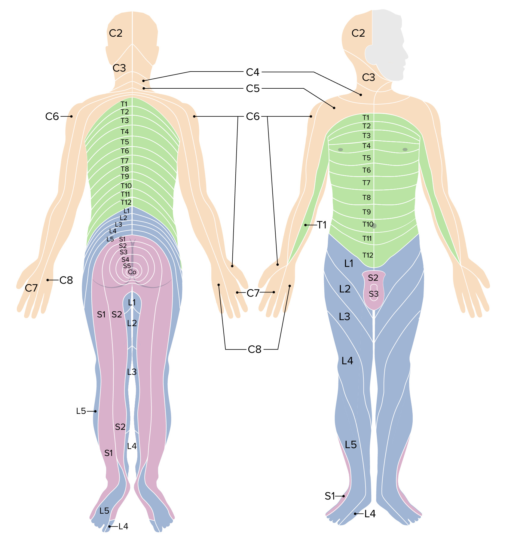
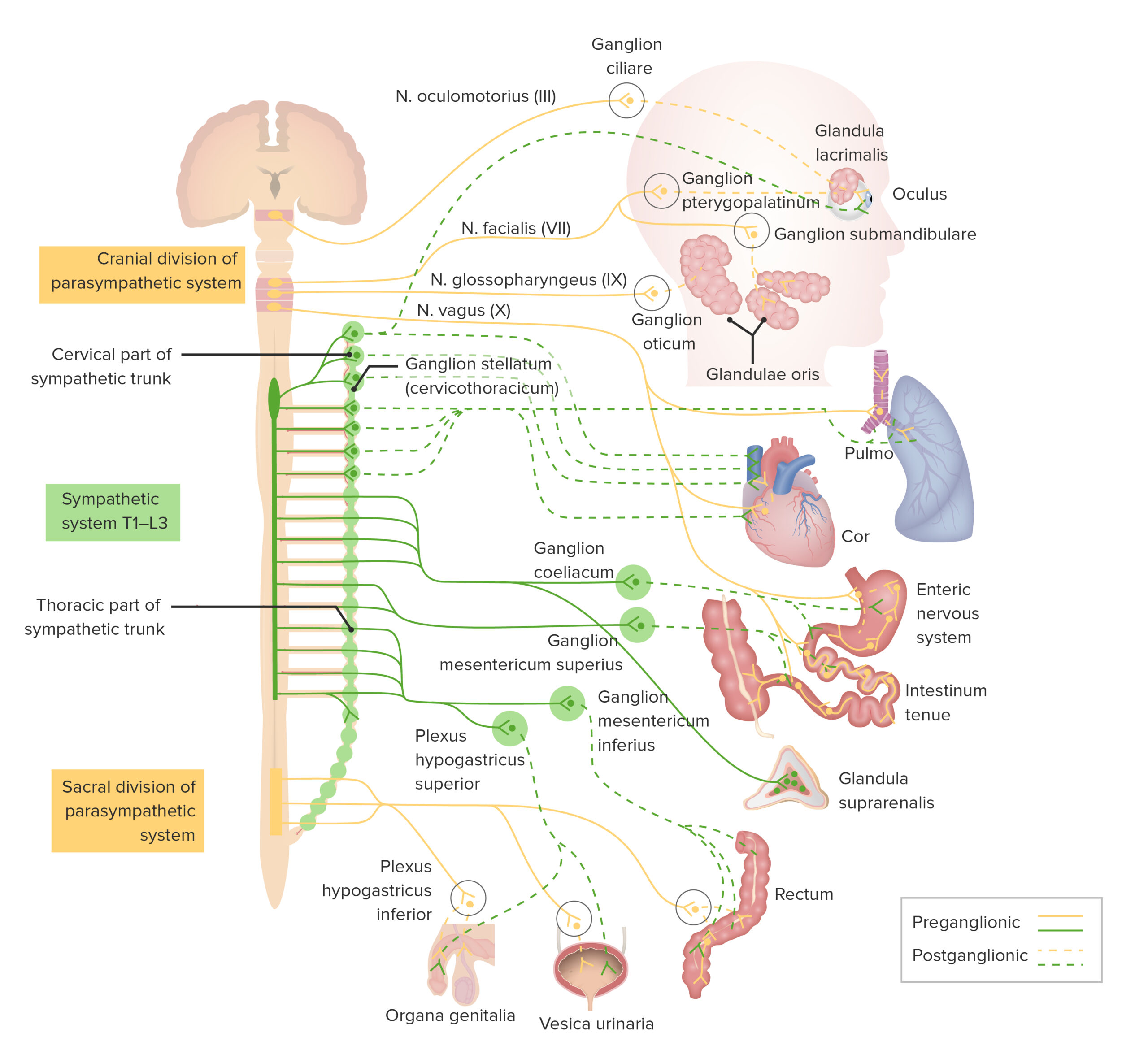
00:00 So let's now have a look at the arrangement and the location of the motor and sensory neurons in the spinal cord. So now it is important for us to make sure we know clearly the locations of sensory and motor neurons. On this diagram, you can see the gray matter and the white matter of the spinal cord. So just get used to this diagram and just have a little view of what sort of information it is going to illustrate. 00:37 I want you to first concentrate on the left-hand part of the figure, the bit that talks about somatic reflexes and we'll talk about the visceral reflexes later on. Picture the peripheral nerve on the outside, and also picture the the ventral horn in the gray matter. And now have a look at the ventral horn. A branch passes from the spinal cord. It is called the ventral root and if you look up top, at the dorsal horn, you can see that this is also a dorsal root and these combine together to form the peripheral nerve. Now, the ventral horn if you look very carefully houses the cell body of the motor neuron, the somatic motor neuron. 01:43 And that neuron has an axon that passes out of the ventral root, travels down the peripheral nerve and innervates the muscle. In this case, it is innervating a finger muscle in response to a finger prick. So that really summarizes the location of a somatic motor neuron, an efferent neuron, exiting the spinal cord. And notice there's only one neuron involved. 02:14 Now picture also on this image the right hand side, the visceral reflex and we will describe now the somatic efferent visceral neuron pathway, the autonomic pathway. Motor neurons that are going to innervate smooth muscle and components that we are not conscious about and again remember that I said two neurons were involved. Well remember that I described the lateral horn when we looked at the section through the spinal cord before. So we have labelled it here on this diagram. Make sure you can recognize that lateral horn. And in that lateral horn is going to be the very first neuron, neuron number 1 and its called the preganglionic neuron. 03:13 Preganglionic because it travels to a ganglion, a group of cell bodies and interacts with the second order neuron, the postganglionic neuron. And in this case if you look very carefully you can see the axon in the diagram moving its way out of the ventral root as well, travelling out of the spinal cord and it travels to this structure labelled called a prevertebral ganglion. 03:44 This prevertebral ganglion is really just outside the spinal cord and I will talk about this in more detail later on. Well, that is where the postganglionic neuron is located and it then travels or its axon then travels and innervates the smooth muscle. So it is important that you understand these two neuron pathway to do with the autonomic nervous system. 04:17 Sympathetic or parasympathetic, follows the same sort of pathway. It is just as we will see later on that location of the ganglion that has the postganglionic neuron is located in the different place, whether it is sympathetic or whether it is parasympathetic. And there is a paraventral or paravertebral ganglion shown there. It does not matter at this stage. What the difference between these two ganglia are? Well let's have a look now at sensory neurons. Where they are located? Have a look again at the peripheral nerve, familiarize yourself again on the somatic reflex side of this diagram and have a look at the dorsal root ganglion labelled there. 05:10 That is where the cell body of the sensory neuron is located, and notice its axon travels from the periphery, from the fingertip picking up the sensation of pain probably from the finger prick, and that pain travels along the axon into the dorsal root ganglion, and travels further on past the cell body of that sensory neuron, into the dorsal root and into the dorsal horn. And there it can interact to the motor neuron, in the ventral horn, by simple interneuron and create the reflex out. So when you prick your finger, sensory information goes in through the dorsal root via the sensory neuron, interacts very quickly with the motor neuron from the ventral horn, and then that motor neuron innervates skeletal muscles to remove your finger. 06:12 Well let's have a look at the visceral sensory neurons. Only one neuron is involved in this situation. Again the dorsal root ganglion shown here helds its sensory neurons. 06:30 All sensory neurons are held in these dorsal root ganglia or in some ganglia in the head to do with sensory neurons carrying in with cranial nerves. And have a look at the visceral sensory neuron, it is detecting information about perhaps the contraction of the wall of the gut, and carrying that information all the way in, through the ganglia. It does not synapse with any neuron. They are all the way in through the dorsal root ganglion and then through the dorsal root just like any other sensory or somatic sensory neuron. That is the pathway then of the visceral sensory neuron.
About the Lecture
The lecture Arrangement and Locations of Motor and Sensory Neurons by Geoffrey Meyer, PhD is from the course Nerve Tissue.
Included Quiz Questions
Sensory information enters the spinal cord via which of the following routes?
- Dorsal roots in the dorsal horns
- Dorsal roots in the ventral horns
- Ventral roots in the ventral horns
- Ventral root in the dorsal horns
- Dorsal roots in the lateral horns
Which of the following reflexes involves an interneuron?
- Pain withdrawal reflex
- Brachioradialis reflex
- Achilles reflex
- Patellar reflex
Author of lecture Arrangement and Locations of Motor and Sensory Neurons

Geoffrey Meyer, PhD
Customer reviews
5,0 of 5 stars
| 5 Stars |
|
1 |
| 4 Stars |
|
0 |
| 3 Stars |
|
0 |
| 2 Stars |
|
0 |
| 1 Star |
|
0 |
Best diagram and explanation ever received. None of my teachers could explain so clear this concept. Thanks Dr. Meyer
