Playlist
Show Playlist
Hide Playlist
Anterior Triangle of the Neck

-
Slides Anatomy Anterior Triangle of the Neck.pdf
-
Reference List Anatomy.pdf
-
Download Lecture Overview
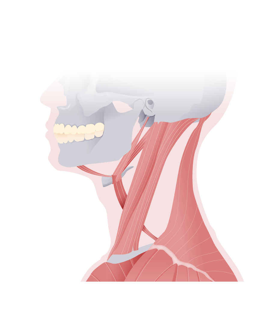
00:01 Next, we're going to look at some anatomic areas that we call the triangles of the neck. 00:07 Now this is a way to divide the neck area to make communication easier when we're talking about physical exam findings. 00:15 For example, location of lymph nodes. 00:18 We'll start by looking at the anterior triangle, which we have a left and a right on either side of the midline. 00:27 The boundaries of this interior triangle are the anterior border of the sternocleidomastoid muscle, the inferior edge of the mandible, and the midline. 00:42 Since that's a relatively big area, sometimes divided further into smaller triangles. 00:51 For example, we have the submandibular, segmental, carotid, and muscular triangles. 00:59 The submandibular triangle is the area that's beneath the mandible, as well as in between the anterior and posterior belly of the digastric muscle. 01:11 The carotid triangle is bounded by the superior belly of the omohyoid, the stylohyoid and posterior belly of the digastric and the anterior edge of the sternocleidomastoid. 01:26 The submental triangle, submental means under the chin, is bounded by the anterior belly of the digastric. 01:34 the hyoid bone in the midline. 01:39 As for the muscular triangle, it's bounded by the hyoid bone, the superior belly of the omohyoid, the sternocleidomastoid and again, the midline. 01:53 Let's start by looking at the muscles of the anterior triangle. 01:57 Using the hyoid bone as our reference. 02:00 We have a group of muscles above called the suprahyoid muscles in a group below called the infrahyoid muscles. 02:08 They also look strapped like. 02:09 So sometimes the infrahyoid muscles are called the strap muscles. 02:15 In terms of the infrahyoid muscles, which we'll focus on in the neck area. 02:20 We have the sternohyoid, the omohyoid, and then deep to the sternohyoid. 02:27 we have the thyrohyoid and the sternothyroid. 02:33 Let's start by looking at the sternohyoid. 02:37 The name is very descriptive because we see it attaches to the posterior aspect of the sternoclavicular joint and travels up to the body of the hyoid. 02:47 hence its name sternohyoid. 02:50 Next we see the omohyoid which is also descriptive. 02:55 because we see it attaches to the superior border of the scapula and omo actually is a word that means shoulder and then it also of course attaches to the hyoid. 03:07 The sternohyoid and omohyoid act to depress the hyoid which is very important in the act of swallowing. 03:17 If we look deep to the sternohyoid we find a shorter muscle called the thyrohyoid which is attaching from the thyroid cartilage up to the greater horn of the hyoid hence its name sternohyoid. It also acts to depress the hyoid. 03:33 Similarly, we have a sternothyroid, which is attaching from the maneuvering of the sternum up to the thyroid cartilage hence its name. 03:44 It also acts to depress the thyroid downward. 03:50 In terms of innervation, the anterior rami of C1, 2, and 3 form this loop like structure called the ansa cervicalis which innovates these infrahyoid or strap muscles with the exception of thyrohyoid. It's innervated by just C1. 04:08 Next we'll look at the jugular veins. 04:12 Here we have the anterior jugular vein or veins. 04:15 There's quite a bit of variability with this set of veins. 04:19 We see that we have the external jugular vein on the external or outer surface of the sternocleidomastoid and the anterior jugular veins are generally going to drain into that external jugular vein, although again, there's quite a bit of variation. 04:38 And here we see the sternohyoid. The sternothyroid, deep to it. 04:43 The omohyoid heading out laterally and the internal jugular vein. 04:49 We also find the ansa cervicalis in this area in close association with the internal jugular.
About the Lecture
The lecture Anterior Triangle of the Neck by Darren Salmi, MD, MS is from the course Neck Anatomy.
Included Quiz Questions
What is the lateral boundary of the anterior triangle?
- Anterior border of the sternocleidomastoid
- Lateral border of the sternocleidomastoid
- Inferior border of the mandible
- Superior border of the mandible
- Midline of the neck
What is another term for the strap muscles?
- Infrahyoid muscles
- Suprahyoid muscles
- Hyoid muscles
- Sternocleidomastoid
- Posterior belly of digastric muscle
What is the origin of the sternohyoid muscle?
- Posterior aspect of the sternoclavicular joint
- Anterior aspect of the sternoclavicular joint
- Body of the hyoid bone
- Neck of the hyoid bone
- Lateral aspect of the sternoclavicular joint
What is the function of the omohyoid?
- Depressing the hyoid bone after swallowing
- Elevating the hyoid bone after swallowing
- Rotating the hyoid bone after swallowing
- Extending the neck
- Flexing the neck
Author of lecture Anterior Triangle of the Neck

Darren Salmi, MD, MS
Customer reviews
5,0 of 5 stars
| 5 Stars |
|
5 |
| 4 Stars |
|
0 |
| 3 Stars |
|
0 |
| 2 Stars |
|
0 |
| 1 Star |
|
0 |
