Playlist
Show Playlist
Hide Playlist
Ankle and Foot Pain

-
Slides FootandAnkle AcuteCare.pdf
-
Download Lecture Overview
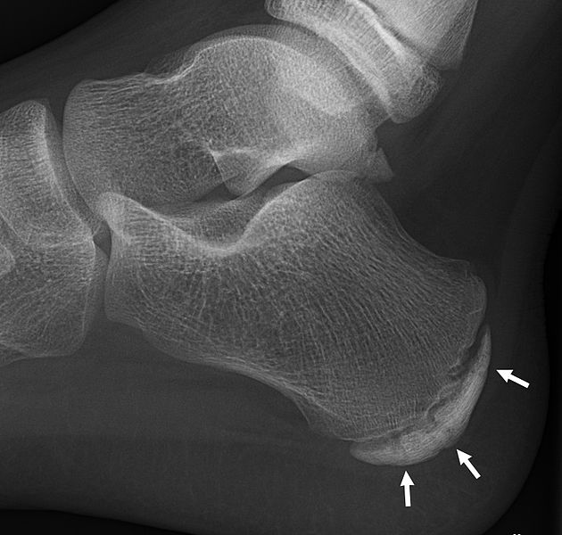
00:01 So, let's talk about ankle and foot pain together, another commonly seen musculoskeletal problem. 00:08 And we’ll start with the approach to the patient with ankle and foot pain. 00:11 And it begins with looking at the acuity of pain. 00:14 Because ankle and foot, I see patients, of course, with acute injuries, lot of sprains and strains, but also who have chronic and debilitating conditions. 00:25 And, of course, they have a precipitating event, the most common being an inversion injury of the ankle, leading to a sprain. 00:32 You’re going to want to record that. 00:35 And that’s why getting a prior history of injury is really important. 00:38 A previous history of sprains begets more sprains. 00:43 Many patients might have conditions that more chronic that relapse and remit over time, and so therefore, getting a prior history of injury. 00:52 And just how serious was it? Did they recover from their ankle sprain and was it always lax and never quite recovered all the way? What kind of activities can they do now? So, a functional assessment is very, very important. 01:04 And then very importantly, more so for my patients with chronic injuries, what kind of footwear are they using? Are they really protecting their foot and giving it the padding and support that it needs to function? So, let's start with something that I think is going to be very germane to the USMLE exam, and that’s the Ottawa ankle and foot rules. 01:26 These are specific rules to help rule out fractures of the foot and ankle. 01:30 The nice thing is they are just like a good screening test. 01:33 They’ve got a very high sensitivity. 01:35 They don't have the best specificity, but what happens, therefore, is that when they’re negative, you can feel fairly reassured that there is no x-ray needed. 01:45 So, the ankle rules start with the fact that the patient cannot walk more than four steps after the injury. 01:51 So, they have to be able to ambulate a little bit. 01:54 And here, what you’re really looking for is, on exam, tenderness over the posterior edge and/or the tip of both the lateral and the medial malleolus. 02:04 Now, so the individuals who suffer an ankle injury, but can walk immediately afterwards and don't have those signs of tenderness on the lateral medial malleoli are therefore very unlikely to have a risk of fracture, and don't need x-rays. 02:19 And saving people x-rays saves them time, saves them exposure to radiation. 02:24 For the foot, it’s similar. 02:25 They have to be able to walk right after the injury. 02:29 The areas to check anatomically on your exam are the head of the fifth metatarsal, so that’s in the mid-foot lateral. 02:36 and then up just at the very top of the foot, over the navicular bone in the midfoot as well. 02:42 No tenderness in those areas, ability to walk after the injury, no need for x-rays in those patients. 02:50 All right. 02:50 So, let's bring the Ottawa ankle and foot rules to life to help understand the anatomic sites to palpate before ruling out fracture. 02:57 And, whoa, is that a really good-looking foot? Whoever’s foot this is is in the wrong business if he is in medicine. 03:05 He should be a foot model. 03:06 Let's start showing the ankle areas for palpation. 03:10 So, that involves the lateral and the medial malleolus from the tip, about 6 cm up the posterior bone of the tibia. 03:20 Like so. 03:21 And so, the anterior side, not so much. 03:24 So, if there is tenderness up here, that's okay because that is a site of a lot of ankle sprains, but it's really along the tip and the posterior side laterally. 03:34 And the same thing medially. 03:35 Tip, lateral side, about that far up. 03:39 That's for the ankle. 03:41 For the foot itself, the base of the fifth metatarsal here, so way lateral, and then over the navicular bone, about here. 03:50 If these areas are not tender, you can feel comfortable that the patient does not have a foot or ankle fracture, and therefore, no x-ray is needed, saving a lot of unnecessary x-rays. 04:03 Just mentioning some other types of tendinopathies. 04:05 Posterior tibial tendon dysfunction, that tendon tends to run right under the foot, but the pain tends be felt more medially. 04:13 It can be misdiagnosed as a medial ankle sprain. 04:16 And left unattended, it can promote flat foot over time. 04:21 On examination, the best way to draw it out is active eversion with dorsiflexion against resistance at the same time. 04:29 And we’ll talk more about this other condition, Achilles tendinopathy, but this oftentimes, you'll find pain, maybe some swelling over the tendon, and it’s often above the insertion of the calcaneus, but you can get a tendinopathy that’s closer to the calcaneus itself as well. 04:44 And it’s very much associated with a change in the intensity of training. 04:48 So, those weekend warriors who get fired up and go out and run 5 miles when they haven’t run in a long time, that's where you see cases of Achilles tendinopathy. 04:58 Plantar fasciitis is one of the more common foot problems that I see in my patients. 05:04 And this one is not related to feeling pain at the end of an activity. 05:09 It's actually worst in the morning. 05:11 Those first steps can be really painful over the plantar foot. 05:14 Similarly, after a prolonged sit. 05:16 A physical examination, very important for these patients. 05:20 And you could see the percentages of patients with plantar fasciitis with particularly tenderness at different points in the plantar fascia. 05:27 I think the main thing that I would note is that the tenderness is usually more medial than lateral. 05:34 So, as you can see, it’s 14% on the midfoot, but right where that 42% is. 05:40 that's where I find most of my patients with plantar fasciitis are most tender. 05:45 I think another thing that's really important to note about this condition is that if the patient has typical symptoms and they have the tenderness in the right areas, very rarely do you need to worry about imaging. 05:56 Many of these patients do have a calcaneal spur on lateral imaging of the calcaneus, but that's not actually related to their fasciitis and the pain. 06:06 So, therefore, you can discard it. 06:07 You don't need to do imaging on those patients.
About the Lecture
The lecture Ankle and Foot Pain by Charles Vega, MD is from the course Acute Care. It contains the following chapters:
- Ankle and Foot Pain
- Specific Ankle/Foot Tendinopathies
Included Quiz Questions
According the the Ottawa ankle rules, which of the following is an indication for ankle X-ray in a patient with pain in the malleolar zone?
- An inability to bear weight both immediately and in the emergency department for four steps
- The patient does not need x-ray imaging for complete evaluation
- Bone tenderness along the proximal 5 cm of the posterior edge of the fibula
- Bone tenderness along the proximal 6 cm of the posterior edge of the tibia
- Bone tenderness at the base of the fifth metatarsal
Which of the following is the most common metatarsal bone that sustains fracture?
- 5th
- 1st
- 2nd
- 3rd
- 4th
An athlete presents with pain 4 cm above the posterior calcaneus after abruptly intensifying his training. Physical examination shows tenderness over the same point. Which of the following is the most likely diagnosis?
- Achilles tendinopathy
- Plantar fasciitis
- Jones fracture
- Medial malleolus fracture
- Navicular bone fracture
Author of lecture Ankle and Foot Pain

Charles Vega, MD
Customer reviews
4,0 of 5 stars
| 5 Stars |
|
0 |
| 4 Stars |
|
1 |
| 3 Stars |
|
0 |
| 2 Stars |
|
0 |
| 1 Star |
|
0 |
Would be better if also included: Hallux valgus,varus,rigidus.
