Topography of the Lungs – Lungs, mediastinum and cardiac valves
About the Lecture
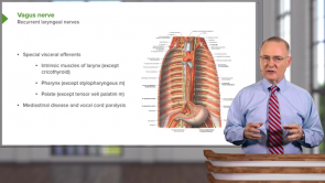
The lecture Topography of the Lungs – Lungs, mediastinum and cardiac valves by Craig Canby, PhD is from the course Thoracic Viscera with Dr. Canby.
Included Quiz Questions
At which vertebral level is the apex of the lung?
- C7
- C2
- C4
- C5
- C3
Which rib correlates to the inferior aspect of the lung along the midclavicular line?
- 6th rib
- 10th rib
- 8th rib
- 12th rib
- 4th rib
![Anatomy [Archive]](https://assets-cdn1.lecturio.de/lecture_collection/image_medium/87992_1693919964.png)
Author of lecture Topography of the Lungs – Lungs, mediastinum and cardiac valves

Craig Canby, PhD
Customer reviews
5,0 of 5 stars
| 5 Stars |
|
1 |
| 4 Stars |
|
0 |
| 3 Stars |
|
0 |
| 2 Stars |
|
0 |
| 1 Star |
|
0 |
Simple, easy to understand
By Nor Syazwani S. on 27. December 2018 for Topography of the Lungs – Lungs, mediastinum and cardiac valves
Short, brief, simple and very informative in just one lecture.
Quiz Overview
wrong
right
open
