Playlist
Show Playlist
Hide Playlist
Visual Processing – Vision (PSY, BIO)


-
Slides Vision SensingtheEnvironment.pdf
-
Download Lecture Overview
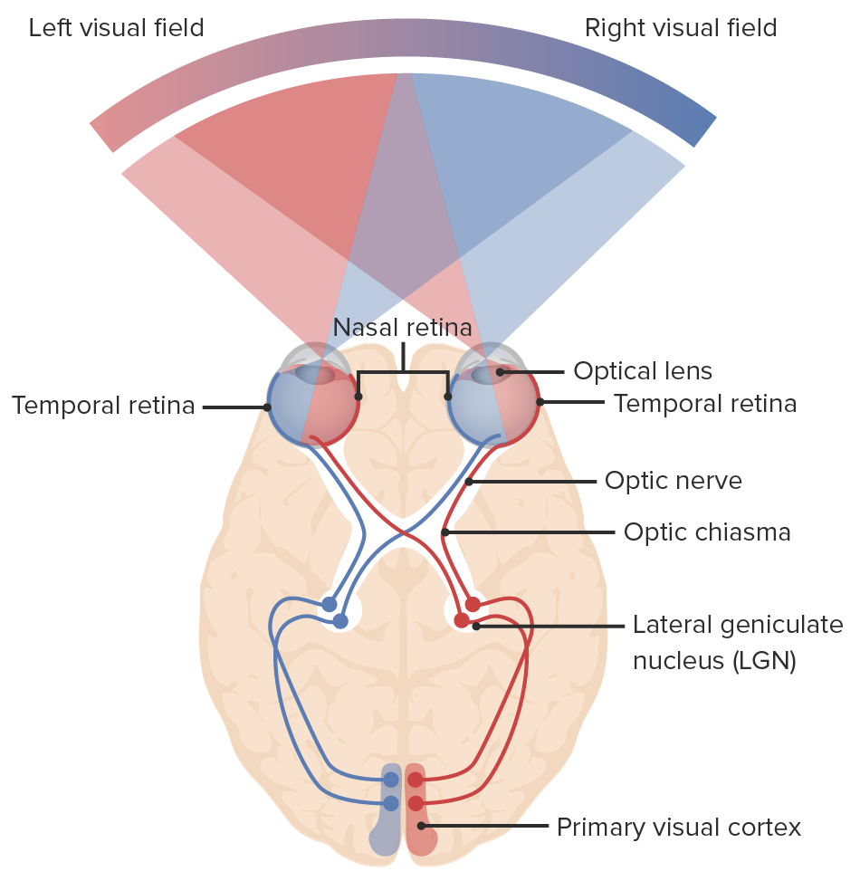
00:01 We’re going to get into now that the light has come in, it’s being detected by the eyes and it’s going to the brain. 00:07 We need to somehow process that information. 00:09 And this is in itself a fairly complicated and robust thing to understand. 00:14 So you’re going to have to probably spend some time looking at this figure that you see here and familiarize yourself with the structures and the way light goes in. 00:22 So a couple of key things to realize, you know we have binocular visions. 00:26 Two eyes, okay? So each eye actually detects a bit from both visual fields. 00:31 So we can have two visual fields. 00:33 So imagine this. 00:34 If you were to take the world and you were to cut it in half, you would have your left visual field and your right visual field. 00:40 Now within each visual field, you’ll have two more hemispheres. 00:44 Okay? And information from both sides of the world go into both eyes bilaterally. 00:51 Okay? So that’s a lot of crisscrossing happening. 00:53 So we’ll walk through a couple of examples and it will start to make sense. 00:56 Once it enters the eye, it crosses yet again. 01:00 And it is processed by the opposing side of the brain. 01:03 And that’s done in the primary visual cortex which is found in the occipital lobe. 01:07 Okay? So the right and left visual fields are processes -- are processed contralaterally in the visual cortex. 01:13 What we’re saying is what you see in the right visual field is processed by the left and vice versa. 01:19 Nerves cross at the optic chiasm. 01:22 So if you look at this diagram, it’s where the tracks are coming in passed the eye and that you see them crossing before they enter the actual lobe of the brain. 01:30 That is called optic chiasm as it’s labeled. 01:33 And it goes through the lateral geniculate nucleus, the LGN. 01:37 The eye is further divided by field, like I’ve mentioned, and visual information is processed ultimately at the primary visual cortex. 01:45 So what we’ll do is we’ll look at the best way we do to understand these types of things is to look at damage, right? We love damage. 01:52 So let’s see what happens when there’s damage within the visual pathway. 01:56 So here’s another orientation of the exact the same diagram, and I want you to note the left eye, the right eye. 02:01 Now let’s focus on the left eye. 02:03 You’ll notice two colors. 02:04 There’s a pinkish color and kind of a bluish color. 02:07 Now on the left eye, the left eye will actually get information from both fields, left field, right field. 02:13 And if you look at the sort of the left eye, you have this blue track and it crosses the optic chiasm and it ends up on the right side. 02:22 So we call that the nasal passage because it’s closes to the nose and you have one on either side. 02:28 You also have, on the outside, you have the optic nerve that’s going and it’s going to stay on the same side of the brain. 02:35 So we get information coming to both sides of the brain. 02:39 But what happens in terms of processing is the left side of the brain will acquire all the information coming from all the right fields but coming from both eyes. 02:48 Okay? So just again, follow the colors. 02:50 If you look at the left eye, you see this pink color coming down and it does not cross the optic chiasm, it actually stays on the same side, and you have information on the blue, staying on the blue side but then you also have crossing. 03:03 Okay? So, let’s take a look at lesions to the right optic nerve. 03:08 So where I have -- where I’ve put the number one is where I’m going to go in and cut that nerve. 03:13 I want you to tell me what do you think is going to happen to your view. 03:17 What are you going to be able to see? So, So I have vision and blind. 03:22 So if it’s filled in black, that means it’s blind. 03:24 And if it’s white, it’s vision. 03:26 You tell me what do you think you’ll see. 03:28 Just take a moment, mull it over. 03:32 Okay. So if you picked complete blindness in the right field, in the right eye, that’s right. 03:38 So if you just follow the lines of where the lesion is made, you can see that that information won’t go to the appropriate lobe where it can get processed. 03:44 Let’s take a look another one that might help. 03:46 Lesion of the midline of the optic chiasm. 03:48 So now what we’re doing is we’re actually cutting right at this midline and so information can’t cross. 03:55 Where are you going to have vision? Where are you going to have blindness? On the outside, you have blindness. 04:01 On the midline, you have vision, and it’s because that we’ve cut that midline chiasm. 04:06 Let’s do one more. 04:09 We have now a lesion at the right side passed the chiasm. 04:17 What do you think you’ll have in terms of vision and blindness? So follow the lines and look at where that light will continue to follow. 04:25 There you go. 04:26 So we have a detriment, a vision for your left visual field -- sorry, the left hemisphere of the field while maintaining your right based on the damage that we have just prior to the lateral geniculate nucleus. 04:38 Okay? So, feature detection and parallel processing. 04:42 Now when we are talking about how the signal actually gets detected and then we establish the different features, that’s really important. 04:49 Again, like sound, vision is detecting a lot of different attributes, things like, again, we don’t really think about. 04:55 So it includes certain things like color, right? We know that’s mediated by the cone cells, but their shape, the lines, the edges, that is done by a specific type of cell called the parvocellular cell. 05:05 And we have movement, which is telling us, you know, something is moving. 05:09 That’s magnocellular cells. 05:11 So these are just three attributes that I’m talking about, but there’s even more than this. 05:15 And all of this happens in parallel. 05:18 So we call that parallel that processing. 05:19 Okay? So I’m going to highlight two of these specific cells. 05:22 It’s important that we know these. 05:24 And that’s the parvocellular cells and the magnocellular cells. 05:26 It’s also called p-cells, m-cells. 05:30 They’re both located in the lateral geniculate nucleus and they both have specific roles in terms of vision, but one is greater spatial acuity or spatial resolution and the other is lower spatial resolution. 05:42 One is lower temporal resolution because it’s not really detecting movement. 05:46 While the one -- the m-cells that detect movement, they want to be good at time. 05:51 One is sensitive to color and one has large fast-conduction neurons because again, it’s trying to detect speed, because it’s trying to detect time. 05:59 You want them to be fast. 06:00 Okay. So in terms of conduction and neurosignals, the larger something is, typically, the faster it is and it’s also in terms of myelination. 06:10 So highly myelinated, really large, it’s going to be really fast. 06:15 Now, again, what are some of the things that we want to talk about? The brain processes all aspect of visual information in parallel. 06:21 So we don’t look at something and first realize that it’s red, oh and then realize that it’s moving, oh and realize that it’s round and realize that it’s actually really close. It’s going to hit me in the face. 06:32 We realize that a red ball is coming, screaming out my face and I need to move, right? So all of that happens simultaneously. 06:39 You know, it has to get integrated at some point. 06:41 So color, emotion, shape, depth The occipital lobe constructs all of these into one overall image. 06:47 So we call that a holistic image. 06:49 It integrates all that information. 06:51 There’s also stored information that’s accessed to speed up the processing and the add context. 06:55 So if you know like cherries are red, then you don’t have to reestablish that every time. 07:02 And if you’re thinking that, you know, that you have black hair again, you don’t need to process that every single time. 07:09 So by storing these concepts, it allows us to speed things up. 07:15 And this is a very resource intensive process that requires 30% of the energy utilization of your cortex. 07:23 That’s a lot considering the amount of information that you actually process just by interacting with somebody. 07:30 Vision really takes the lion’s share of the resources. 07:33 So we just walked all the way through the different components of the eye. 07:38 We talked about all the different aspects, how we have rods and cone cells, how those are -- the process of phototransduction, and then we finished off on looking in how all this information is processed and detected.
About the Lecture
The lecture Visual Processing – Vision (PSY, BIO) by Tarry Ahuja, PhD is from the course Sensing the Environment.
Included Quiz Questions
What defect would be caused by an optic nerve lesion (pre-optic chiasm)?
- Total blindness of 1 eye
- Temporal hemianopia
- Hemianopia with macular sparing
- Contralateral hemianopia
- Quadrantanopia
What is the order of the visual pathway?
- Optic nerve, optic chiasm, optic tract, lateral geniculate body, optic radiation, and visual cortex
- Optic tract, optic chiasm, lateral geniculate body, and visual cortex
- Optic nerve, optic tract, optic radiation, and visual cortex
- Optic nerve, parvocellular cells, optic radiation, and visual cortex
- Optic nerve, optic tract, optic radiation, optic chiasm, lateral geniculate body, and visual cortex
Which description regarding parvocellular cells is accurate?
- Sensitive to color
- Involve highly myelinated nerves
- Involved in visual resolution
- Located in the medial geniculate body
- Involved in detection of color
Which description regarding feature detection is accurate?
- Uses parallel processing
- Uses the frontal lobe
- Uses 10% of the cortex
- Uses the parietal lobe
- Uses 50% of the cortex
Author of lecture Visual Processing – Vision (PSY, BIO)

Tarry Ahuja, PhD
Customer reviews
5,0 of 5 stars
| 5 Stars |
|
5 |
| 4 Stars |
|
0 |
| 3 Stars |
|
0 |
| 2 Stars |
|
0 |
| 1 Star |
|
0 |
