Playlist
Show Playlist
Hide Playlist
Vestibulo—Ocular Reflex (VOR)

-
Slides 10 VestibularSystem 1 BrainAndNervousSystem.pdf
-
Download Lecture Overview
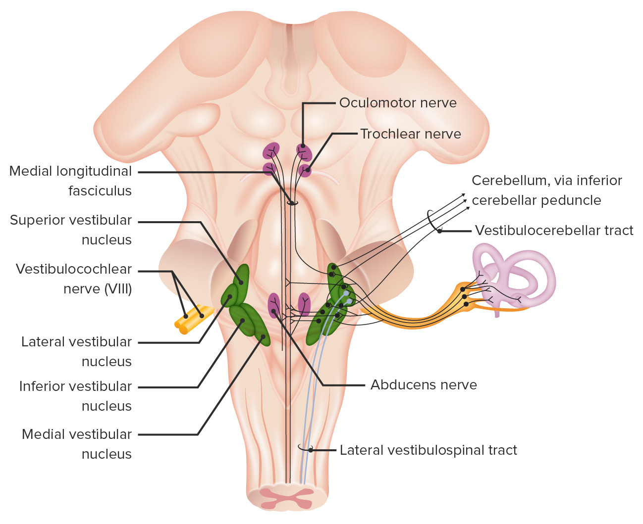
00:00 There is a reflex that’s important for you to understand. This is the vestibulo-ocular reflex, abbreviated V–O–R. 00:11 The vestibulo-ocular reflex, what happens here is if your head moves to the right, for example, and your eyes would remain fixated on the object that you’re initially at, this will allow even as you’re moving your head to the right, for example, that your eyes will remain fixated on the object back to your left. 00:37 So if your head’s moving to the right, your eyes have to move together back to the left to maintain that visual field. This is a reflex that is composed of three neurons. So what is a three neuronal circuit? So let’s explore this. Let’s look at some of the neurons that are involved in this circuitry and look at what muscles have to be activated in order for this to work appropriately. The first thing to set up here is, we have a posterior view. We’re looking at the back of the head of this particular individual. 01:19 We’re also looking at the brain stem and even the spinal cord in a posterior view. So the right nuclei that we see here at multiple levels, these are on the right side as this is the back of the head being on the right. 01:36 Then the nuclei that we see here are associated on the posterior left in this particular view. 01:45 We’re going to have this individual move their head to the right and again the eyes will want to move back to the left. So what circuits have to be activated in order to have this reflex occur appropriately or normally? The first consideration here is that as you move your head, you’re going to disrupt the vestibular hair cells so that they start to fire. So you activate your 1st order afferents in the vestibular ganglion from cranial nerve eight on the right side. 2nd order neurons in the vestibular nucleus will then activate 3rd order neurons in the extraocular nucleus. Then the appropriate extraocular nuclei can activate the appropriate muscles to move the eyes to the left as the head moves to the right. 02:47 So the hair cells in the right semicircular ducts will increase their firing rate in response to head movement to the right. The 1st order afferents in the right vestibular ganglion that houses the cell bodies of these 1st order nerve cells will send the axons out toward the vestibular apparatus as they become activated or stimulated. The extraocular nuclei are next stimulated. First would be the ipsilateral oculomotor nucleus here. Then you’ll activate the contralateral abducens nucleus that we see on the left side. When we think about what has to happen to move the eyes back to the left, again, we’re activating the right oculomotor nucleus. So then, we’ll activate the oculomotor nerve fibers to the right. 03:50 These are going to innervate the medial rectus of the right eye and then cause the eye to move, the right eye to move to the left through a shortening of the medial rectus muscle. The activation of the contralateral abducens nucleus will send action potentials along the length of the abducens nerve fibers to the lateral rectus muscle of the left eye causing it to shorten and to move the left eye to the left. As a result of moving both eyes to the left, the visual field will remain stable even as the head rotates to the right.
About the Lecture
The lecture Vestibulo—Ocular Reflex (VOR) by Craig Canby, PhD is from the course Auditory System and Vestibular System.
Included Quiz Questions
Which of the following structures contains the 1st-order neuron for the vestibulo-ocular reflex?
- Vestibular ganglion
- Celiac ganglion
- Ciliary ganglion
- Otic ganglion
- Spiral ganglion
If the head moves toward the right side, which of the following cranial nerve nuclei will be stimulated?
- Ipsilateral oculomotor and contralateral abducens
- Ipsilateral oculomotor and ipsilateral abducens
- Ipsilateral trochlear and contralateral abducens
- Contralateral oculomotor and ipsilateral abducens
- Ipsilateral oculomotor and contralateral trochlear
For a person with an intact vestibulo-ocular reflex, which of the following statements is most accurate?
- If the head moves left, both eyes will move to the right.
- If the head moves left, both eyes will move to the left.
- If the head moves right, the left eye stays stable, and the right eye will move to the right.
- If the head moves right, both eyes will move to the right.
- If the head moves left, the right eye will move to the right, and the left eye will move to the left.
Author of lecture Vestibulo—Ocular Reflex (VOR)

Craig Canby, PhD
Customer reviews
5,0 of 5 stars
| 5 Stars |
|
5 |
| 4 Stars |
|
0 |
| 3 Stars |
|
0 |
| 2 Stars |
|
0 |
| 1 Star |
|
0 |
