Playlist
Show Playlist
Hide Playlist
Varicocele

-
Slides Introduction Male Repro.pdf
-
Download Lecture Overview
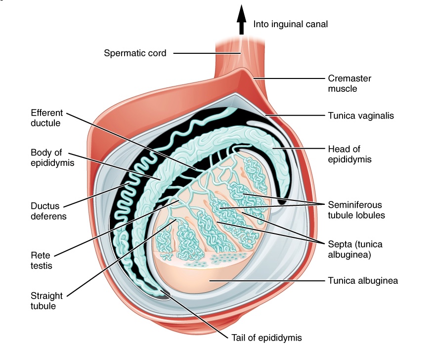
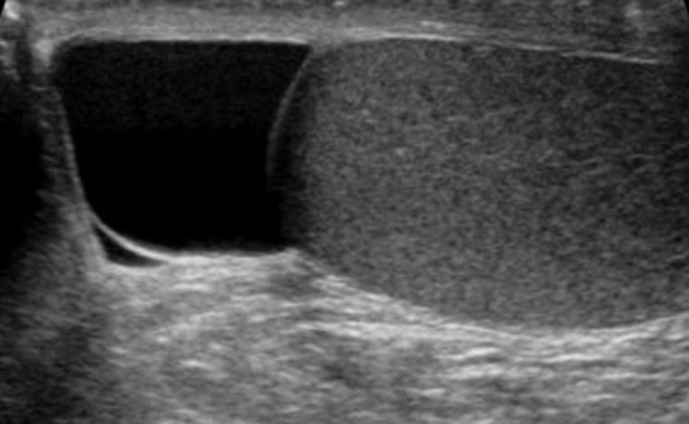
00:02 Our topic here is varicocele. 00:04 What do varices mean to you? Engorgement of your veins. 00:08 Here we have a varicocele. 00:11 What’s happening? Dilation of veins, you must know the anatomy in greater detail here. 00:16 You must know about the pampiniform plexus due to increased venus pressure. 00:21 How does your increased pressure which is then – Well, the veins, what are they doing? That’s important for you to understand. 00:28 Are veins draining or supplying? Draining. 00:31 So if there’s increased pressure in the veins, then you’re having difficulty with the draining. 00:36 So now, what happens? Engorgement, right? Welcome to varicocele. 00:42 Concept first, then you plug in the diagnosis. 00:46 Most common cause of scrotal enlargement is varicocele. 00:50 Most common cause of scrotal enlargement, hence, know everything about varicocele. 00:57 What side? Almost always on the left side due to resistance to flow. 01:02 It’s important that you know the anatomy here. 01:05 The left testicular vein or gonadal vein drains into the left renal vein. 01:10 Can you picture that for me? Does that happen on the right side? No. 01:15 Okay, so pause. 01:18 On the left side, if the left gonadal vein or testicular vein has to drain into the left renal vein first, then it drains into your inferior vena cava. 01:26 My goodness, that’s a pretty long journey. 01:29 And so therefore, if at any point in time there is blockage, then you’re going to have engorgement and enlargement of the scrotum, left side. 01:37 Simple anatomy, clinical – Everything that you do in medicine between you and I or in general by yourself, make sure you give it a clinical tag. 01:45 If you don’t, then I don’t know, just leave it aside and come back to it. 01:50 Acute onset may indicate a thrombotic event. 01:53 Secondary to nephrotic syndrome or even renal cell carcinoma. 01:57 Keep that in mind. 01:58 Interesting? We’ve talked about this. 02:02 Nephrotic syndrome leaves you in what kind of state? Bleeding or hypercoagulable? Good, hypercoagulable. 02:10 Do you see as to how everything that you’ve learned is coming together now? Nephrotic syndrome, your losing antithrombin III. 02:17 Antithrombin III normally knocks out your? If you remember. 02:19 Thrombin. 02:21 So normally, it should control coagulation. 02:25 In nephrotic syndrome, if you remember, of all the coag factors, the one that you have memorized and, if you haven’t, do so. 02:31 Again, now, you lose antithrombin III. 02:36 And you're state of what? Hypercoagulabilty. 02:39 Where? Renal vein. 02:42 And what about renal cell carcinoma? Renal cell carcinoma, once again if it’s the left side, think about metastasis. 02:50 You’ve moved from stage one to stage two. 02:52 You’ve busted through the capsule and now you’re going to metastasize through the renal vein. 02:58 This is metastasis. 03:01 All these cancer cells, slowly moving. 03:04 It’s a gang passing through the renal vein. 03:07 And all of the sudden, on the left side, here comes gonadal. 03:10 Hey, what’s up? But the thing is, it’s a bully. 03:14 It stops the drainage of your gonadal vein So you’d stopped the drainage of your testicular vein or gonadal vein, welcome to varicocele. 03:24 Signs and symptoms that will then help you out to figure out what’s going on with your patient. 03:30 Next, well, if you’re decreasing the drainage, would you call this hyperemia or congestion? Yet another basic concept. 03:38 Congestion. 03:40 So congestion, may result in high temperatures. 03:43 What’s optimum temperature? How about 98.6 degrees Fahrenheit, 37 degrees Celsius? With all these increased heat, there’s every possibility that you may or may then become infertile. 03:56 So far so good. 03:58 Let’s keep going. 03:59 Diagnosis made by standing exam. 04:01 What? Yes, exactly right. 04:04 You can see this on gross examination with your eyes and the scrotum looks like it contains a bag of “worms”. 04:12 Why? Because the veins are engorged. 04:14 Can you picture that? Literally, the scrotum looks like a bag of worms. 04:19 Okay. 04:20 Hence, the standing exam. 04:22 Let’s keep going. 04:23 Inspect the bag of worms that are palpable and observe with? Make sure you know how to investigate properly. 04:29 A Doppler ultrasound. 04:31 It does not transilluminate. 04:33 That’s important. 04:34 It does not, because what’s in your vein? Blood. 04:38 Blood is thick and viscous, it will not transilluminate. 04:42 "Versus what, Dr. Raj?" Hydrocele. 04:46 Be careful. 04:48 They both have the suffix –cele. 04:50 Varicocele, hydrocele. 04:52 You pay attention to the prefix. 04:54 Varico-, blood. 04:56 Hydro-, fluid. 04:58 Which one of these will illuminate? Fluid, hydro-. Not varico- Are we clear? Let’s keep going. 05:05 So it does not transilluminate. 05:06 And management includes – Well, you have to – My goodness, you have to get rid of the congestion. 05:11 So there’s something called varicocelectomy or perhaps something called embolization. 05:16 On a Doppler ultrasound, you will find certain issues and with ultrasound, you would find as you see in the picture here, the bag of worms type of appearance. 05:27 Image shows several what’s known as your anechoic tubule. 05:31 What does that mean to you? Remember this doesn’t appear as being lucent and black, anechoic. 05:36 And the application they call a Doppler imaging in the same patient will show what? Bidirectional flow with anechoic tube. 05:44 Now, do you understand this is in the veins? Should the veins ever -- The fluid and the blood, should it ever move back and forth, back and forth? No. It’s always unidirectional. 05:54 Veins are always draining. 05:57 So if it’s blocked and you see bidirectional flow, that’s a pathology. 06:02 Full picture of varicocele. 06:03 Every single bullet point that I’ve gone through here, incredibly important.
About the Lecture
The lecture Varicocele by Carlo Raj, MD is from the course Male Reproductive System Diseases.
Included Quiz Questions
Acute onset of a varicocele may indicate what pathology?
- A thrombotic event
- Testicular cancer
- Early renal cell carcinoma
- Infertility
- Testicular torsion
Which factor does NOT typically cause an acute varicocele?
- Acute thrombosis of the left popliteal artery
- Left renal cancer
- Retroperitoneal masses
- Nephrotic syndromes
- Hypercoagulable states
Which statement is TRUE regarding the diagnostic findings of a varicocele?
- Color doppler shows bidirectional flow
- A varicocele appears as a hyperechoic tube on ultrasound.
- Transillumination is seen with a varicocele.
- A varicocele feels like a large cyst since it is filled with fluid.
- A varicocele decreases in size with standing.
Author of lecture Varicocele

Carlo Raj, MD
Customer reviews
5,0 of 5 stars
| 5 Stars |
|
1 |
| 4 Stars |
|
0 |
| 3 Stars |
|
0 |
| 2 Stars |
|
0 |
| 1 Star |
|
0 |
1 customer review without text
1 user review without text
