Playlist
Show Playlist
Hide Playlist
Structure and Function of the Eye – Vision (PSY, BIO)
-
Slides Vision SensingtheEnvironment.pdf
-
Download Lecture Overview
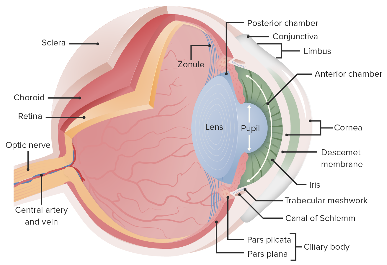
00:01 I got my eye on you. 00:03 Let’s talk about vision. 00:04 We’re going to talk about structure and function of the eye. 00:06 So, the eye is comprised of three layers: the sclera, the choroid, and the retina. 00:13 The sclera is what gives us the white in our eyes. 00:16 That’s the white portion of the eye and that’s the outermost layer. 00:18 Then we have the choroid, which is the darkly colored -- the darkly colored layer and that absorbs all the excess light. 00:26 And then we have the retina which is the one that you hear about the most, and this is the surface where light is actually focused and a lot of that processing happens. 00:33 So we can see all the three layers here. 00:36 And you’ll also notice that the sclera continues to go over the front of the eye and that’s a clear portion of the eye that we’ll talk about in just a sec. 00:43 So the idea here is light is refracted as it passes through the cornea. 00:46 So the outside light coming in goes through to the cornea, which is an extension of the sclera. 00:51 And its job is to actually control where it’s going to go a little bit. 00:56 It enters the anterior chamber. 00:58 So there’s a chamber there that’s filled with a fluid termed aqueous humor. 01:02 You’re going to need to know that term for sure for the MCAT. 01:04 So one way you can try to remember that is aqueous humor, funny, water. 01:09 I don’t know. Sometimes that helps me. 01:10 Sometimes it’s stupid, but I like stupid things. 01:13 So aqueous humor, funny, water. 01:15 Now that, that fluid fills that chamber. 01:19 And what happens is as the light enters, there’s an iris. 01:22 And the iris is the colored part of the eye, that’s what gives us the color of our eye, and it has an opening called the pupil which appears black and that’s where the light is going to enter. 01:33 Now, the size of that pupil, we can control and we can influence. 01:38 So the size of the pupil controls the amount of light which is going to enter the eye. 01:42 And this is mediated by two separate muscles. 01:44 We have the dilator muscle and the sphincter muscle. 01:47 And so the way to remember that, I think we all heard the term before dilation or to open. 01:52 So this is a muscle that would open your eye. 01:54 And then the sphincter, you can think of sphincters in your GI tract or your rectum. 01:59 That makes things smaller. Okay? So dilator muscle opens, sphincter muscle makes small. 02:03 So it’s kind of interesting to note where these are coming from and that we have constriction and we have dilation. 02:12 Now, the constriction is controlled by the parasympathetic system, while the dilation is actually mediated by the sympathetic system. 02:19 If you recall from previous lectures, parasympathetic refers to the rest and digest action, and sympathetic refers to fight-or-flight. 02:28 Now, if you think of the fight-or-flight situation, that’s when you’re aroused and you have a stressor in front of you and you need to make a decision, one of the actual physiological effects that you see in individuals is a dilation of their eyes. 02:41 And you might imagine this because they need to take in as much information and you get this tunnel vision where they’re really focused at the task at hand because that's their stressor. 02:50 So it’s not a coincidence that this is controlled by the sympathetic system while the rest and digest, you’re relaxed, the eyes are droopy, you don’t really need to be focused that much, you see the constriction. 03:02 Okay. 03:03 So, let’s take a look at once the light has entered and passed into that chamber. 03:09 Behind that posterior chamber is the lens which fine-tunes the angle of the incoming light. 03:14 So this is really, really great. 03:16 If you ever played with a microscope in class, you have two knobs; you have your coarse adjustment knob and your fine adjustment knob. 03:23 So we’re now entering the fine adjustment portion. 03:26 Okay? So here what happens is that lens is going to focus the light on a part of the retina, the back of the eye that we wanted to. 03:35 And so we can control. 03:37 We can control the curvature of this lens. 03:40 Now the curvature of the lens is normally convex. 03:42 If it was not convex, we’d be concave. We’d be weird. We’d have weird-looking eyes. 03:47 So it’s actually convex as you can see here. 03:49 And it controls refractive power. 03:53 So you should know that as well. 03:54 And it does that through a certain muscle called the ciliary muscle. 03:58 When the ciliary muscle contracts, this increases the curvature of the lens, thus facilitating near vision Okay? So, here’s a lot here to digest. 04:08 There’s a lot of information, but we’re going to walk through the major important components. 04:12 You know you’re going to probably have to spend some time studying this diagram. 04:16 But for now, we want to appreciate the different layers and the components that make up this diagram. 04:22 So, I want you to notice at the top, we have some structures that look like some rods and some look like different cones. 04:29 Those are photoreceptors called rods and cones that we’re going to look at in a little bit more detail. 04:33 They project to a bunch of neurons, bipolar cells, and we collectively call this other layer interneurons and these will then go on to connect to retinal ganglion cells which we’ll look into more detail. 04:46 And those ultimately exit the eye and go to the brain. 04:49 Now, for the MCAT, you should be well-aware of these different layers. 04:54 We should be aware of the orientation as well. 04:56 And we should be aware of the fact that this is part of the retinal layer of the eye. 05:00 Now, you might think these rods and cones need to detect the light and so lights going to hit them. 05:06 It’s actually flipped. 05:07 The light comes in through this network and hits, you can see the arrow pointing up, that represents the light going in and it actually is as far possible from the rods and cones and makes its way to the rods and cones. 05:22 So, the retina contains two types of electromagnetic receptor cells, photoreceptors called rods and cones. 05:29 The rods and cones synapse with bipolar cells which then synapse with retinal ganglion cells. 05:34 The cones and rods are the most deep layer. 05:38 So light comes from the bottom and they’re at the far end. 05:41 And the axons of the ganglion cells from the optic nerve -- sorry form the optic nerve. 05:46 So all of the ganglion cells coalesce together and create a sort of a bundle of fire. 05:50 Imagine a computer setup, a desk at home with all your printer cables and monitor cables and you bundle them all out and that’s what leaves your desk sort of same idea. 05:59 Now, let’s take a look at a blowup, a small section of the retinal layer and you can see the orientation that I’m talking about now. 06:10 So we can see that we have the rods and cones on the far end and we can have the interior as the retinal layer and we have the interneurons in between and these all interact in synapse with the retinal ganglion cells which go on and form the optic nerve exiting the eye. 06:25 Now, the point on the retina where many axons from the ganglion cells converge to form this optic nerve and that leave the eye, that area is called the optic disk. 06:37 So there’s a spot within your eye because it’s leaving the eye, you won’t have any receptors there. 06:42 And that is what we call the blind spot. 06:45 So we’ve all experienced this before and there’s lot of different test you can do of putting something in front of you and moving your head away and you’ll notice that you can no longer see it. 06:54 If you’ve ever driven a car, you know you have something called your blind spot, and that’s where you have that spot in your eye where you can’t look or it’s a spot that you can’t see anything. 07:02 That’s what they’re referring to. 07:03 So one of the simplest experiments is on a piece of paper draw a black dot or an X. 07:09 And as you move it away, closer move or it farther and closer to your face, you’ll notice the spot where you have that blind spot in terms of distance. 07:18 So the macula is an oval-shaped pigmented area near the center of the retina. 07:23 And you got to imagine a 3D space. 07:26 In the center of that is something called the fovea centralis, which is the largest concentration of cone cells, and this is responsible for central, high-resolution vision. 07:36 And we’re going to get into the difference between rods and cones, but cones are really, really high-resolution cells and they detect color. 07:45 And so this is the spot in the fovea centralis where we have the highest concentration of color vision, which is what we use most of the time, and so that’s where a lot of our light that’s entering the eyes is focused upon.
About the Lecture
The lecture Structure and Function of the Eye – Vision (PSY, BIO) by Tarry Ahuja, PhD is from the course Sensing the Environment.
Included Quiz Questions
Which of the following has the greatest influence on the refractive power of the eye?
- Cornea
- Aqueous humor
- Ciliary muscle
- Dilator muscle of the iris
- Retina
What changes occur in the eye while a person is running away from a bear?
- Contraction of the dilator muscle of the iris
- Activation of the sphincter muscles of the iris
- Increased production of aqueous humor
- Absorption of all excess light by the choroid
- Contraction of the ciliary muscles to focus better
What structures does light pass through in the eye (in anatomical order)?
- Cornea, anterior chamber, pupil, lens, ganglion cell layer of the retina, interneurons, and rods and cones
- Sclera, choroid, and retina
- Retina, choroid, and sclera
- Cornea, anterior chamber, lens, pupil, interneurons, and ganglion cell layer of the retina
- Cornea, aqueous humor, iris, lens, retina, and optic nerve
What structure(s) is/are involved in the refractive power of the eye?
- Cornea and lens
- Cornea only
- Iris
- Lens and retina
- Lens only
What defines the blind spot of the eye?
- The area of the retina without photoreceptors
- The area of the retina with the largest concentration of rods and cones
- The black area in the center of the iris
- The area of the retina where dendrites of ganglion cells converge
- The hyperpigmented area of the retina
Where does the processing of light occur?
- Retina
- Sclera
- Iris
- Lens
- Choroid
Customer reviews
5,0 of 5 stars
| 5 Stars |
|
4 |
| 4 Stars |
|
0 |
| 3 Stars |
|
0 |
| 2 Stars |
|
0 |
| 1 Star |
|
0 |
I enjoy Prof. Tarry's energetic lecturing style and also how he gives out practical examples to facilitate learning. Way to go prof!
VERY high quality lecture. GREAT professor. Clear and concise. Awesome
I am not a pre-med or medical student; physical therapy instead. So far in the first hour of viewing this site I am impressed. Hopefully it will help me achieve my goals. This lecture was outstanding too! I really enjoy the association style teaching Dr. Tarry uses.
very helpfull lectures, ??? ?????? ?? ?? ?????? ????????..???? ???


