Playlist
Show Playlist
Hide Playlist
Structure and Function of the Central Nervous System – Biological Bases of Behavior (PSY, BIO)

-
Slides Biological Basis of Behavior.pdf
-
Download Lecture Overview
00:02 Okay. Now let’s move to our counterpart, which is the central nervous system. 00:05 So like I said, it’s made up of the spinal cord and the brain. 00:08 The brain has three main subdivisions, the hindbrain, midbrain, and forebrain. 00:13 So there are also the older names that have been used, the rhombencephalon, the mesencephalon, and prosencephalon. 00:18 You should probably know those for the MCAT because they might not refer to them as hindbrain. 00:22 They most probably will, but they might also throw around these other name, so you should kind of associate which one goes with which. 00:29 The entire CNS is contained within this sac and it’s bathed in this solution called the cerebrospinal fluid or CSF and it’s basically a salt solution so it’s saline. 00:40 We say it’s a circulating saline solution which provides all the different nutrients, protection, and removes waste. 00:46 So nutrients make sense because it’s bathed in stuff and it has all the ions, the sodium, the potassium, the calcium, and all those things that it needs. 00:52 And it provides protection because the brain actually sits floating inside the skull, inside different layers of membrane and this cerebrospinal fluid. 01:06 So the analogy I like to use is think of maybe a large container of soup or something like a metal tin and if inside that you put a water balloon and then you fill that up with a little bit of water and put the lid back on. 01:21 So that would sort of represent the skull and brain with the outside of that hard tin being the skull, then you have your brain which is the water balloon, and then you have water and that kind of allows it to float around. 01:31 And you want that because if there was not CSF, you would have your very tender, soft, malleable brain tissue against this rigid, hard skull and just walking around or dancing, you basically would be causing major brain trauma, which you don’t want to be doing. 01:48 So by having this kind of floating system, it allows it that buffer and that ability to deal with a lot of the movement and impact that we deal with. 01:59 Here’s the basic structure, again, not a lot of components. 02:02 We have the brain, we have the spinal cord, and then we have everything else, which is the peripheral nervous system, we have the ganglion cells, and we have all the nerves that exit the spine going to all the different effectors and getting sensory information as well that goes up to the spinal cord -- up through the spinal cord to the central nervous system. 02:19 So easy divide. Brain, spinal cord, CNS; everything else is PNS. 02:26 Here’s a nice blowup of the spinal cord. 02:28 It shows that it’s actually a stacking of different bone segments and each one is separated by some tissue and cartilage and there’s a cavity that allows these nerves to go in and to go up to the spinal cord. 02:43 So it’s connected to the brain and is protected by the CSF as well, so it also has the same fluid and has a vertebral column. 02:52 This is the pathway for sensory data to go to the brain and it allows for integration and processing up at the brain. 02:59 There’s some integration and processing that actually happens in the spinal cord too. 03:03 So as things come in, they synapse, and you’re starting to see some integration happen right on the spot. 03:09 And it’s also responsible for simplest spinal reflexes like the muscle stretch reflex which we talked about. 03:14 So it prevents it from having to go all the way up to the brain. 03:16 It can happen right there at the synapses happening within the spinal cord. 03:23 Now let’s take a look at some of the structures in each of those different regions that we talked about. 03:27 We’ll start with the hindbrain. 03:28 So first off you have the medulla oblongata. 03:30 This is a spot that relays information between the different areas of the brain. 03:35 So it’s sort of like a major hub. 03:38 It regulates vital autonomic functions including blood pressure, digestive function, vomiting. 03:44 So again, these are things that you might not necessarily be thinking about. 03:47 And kind of a simple rule of thumb that we have for the brain is the deeper you are in the brain, sort of the more rudimentary or simple the function, and as you peel that onion and you go on the outside -- actually, not peel the onion, but add layers to the onion and move farther away, the more sort of complex and higher level the function is. 04:08 So the very -- the base of the brain being simple stuff, and the very outside or the cortical layers is where we do a lot of our executive function and higher level thinking. 04:17 So the pons is a connection point between the brainstem and the cerebellum and this coordinates movement and balance. 04:23 So we’re not going to spend tons of time going through each of these because simply put you need to know these structures and you need to know their basic function for the exam. 04:32 The cerebellum is also known as little brain and sort of basically because of its function and its location. 04:40 So we say it’s an integrating center where complex movements are coordinated. 04:45 The midbrain now is a relay for visual and auditory information. 04:51 So this is where a lot of that input is now coming in. 04:53 And it also contains the RAS, which has come up at other lectures as the reticular activating system, which is responsible for arousal and wakefulness. 05:01 And the link I always tell people if you don’t remember think of arousal or being activated and this is called the reticular activating system or RAS. 05:11 The brainstem refers to the midbrain along with the medulla and pons. 05:15 So you’ll also hear that term brainstem, I mean I myself have been using it already and that’s what we’re referring to actually, is the midbrain and the medulla and the pons. 05:24 Now the forebrain includes the diencephalon and telencephalon. 05:28 And the diencephalon includes structures like the thalamus and the hypothalamus. 05:32 The thalamus contains relay and processing centers for sensory information, and the hypothalamus contains centers for controlling emotions, autonomic functions, and a major role in hormone production and release. 05:43 So quite a bit of stuff they’re doing, this is kind of advanced stuff, so now layering an emotion and processing actual sensory information. 05:51 This is sort of higher level stuff. 05:55 Here’s a nice image looking at all the different components of the central nervous system as a whole and there’s a few that I want to highlight. 06:05 One, the locations of the structures that we’ve already talked about, so the hypothalamus, the midbrain, the cerebellum, the brainstem. 06:13 I want you to also notice how the spinal cord comes up and then is connected to the brainstem and it’s kind of at the base of the brain. 06:21 And we have something called the corpus callosum. 06:24 Corpus callosum, this is also very important. 06:26 And this is a bundle of fibers that connects the two hemispheres of the brain. 06:33 So we haven’t gotten there yet, but basically the brain is made up of two halves. 06:36 I’m sure you’ve heard of that before, left brain, right brain, but there’s actually a physical clear marcation between the two sides of the brain. 06:46 The two hemispheres are connected, one of the connections is through this corpus callosum. 06:51 So here’s an image looking at, you know, an animal model of the brain and you can see as you spread the hemispheres apart, you can clearly see the corpus callosum. 07:02 So it connects the two cerebral hemispheres. 07:05 In this diagram you also can see the location of the cerebellum, as well. 07:12 Now the two sides have some differences. 07:15 So generally speaking, both hemispheres have similar functions, but there’s a little bit of differentiation in each. 07:21 So for example, at the rear of the brain at the occipital lobe, we know that that lobe is primarily dealing with visual function. 07:29 Now, the left and right side of the occipital love both deal with visual function, but they might control and modulate and mediate different aspects of vision. 07:38 So the hemispheres are bilateral, meaning two sides, left and right. 07:43 The left side of the brain controls the motor function of the right side, and the opposite holds true. 07:49 So when I’m moving right hand I’m actually using the left side of my brain, and when I’m using my left hand I’m using the right side of my brain. 07:56 So information is crossed. 07:59 The left hemisphere is generally responsible for speech, while the right hemisphere is responsible for visual-spatial reasoning and for interacting with things like music, a little bit more of the artsy side, okay? So here are the lobes, you’ve seen these before. 08:15 You should be very familiar with their location and their names. 08:19 So the frontal lobe, top of the hat, it’s nice to meet you and you’ll remember that because it’s at the front of your brain, okay? Then we have the occipital lobe which is at the back of the brain and this is the part of the brain that deals with visual function. 08:32 And then we have the parietal lobe which is kind of in between the two. 08:35 And then the temporal lobe if you want to remember is where your temples are, okay? So you should know their locations. 08:41 Now the frontal lobe initiates all voluntary movement, complex reasoning, problem solving. 08:47 So the prefrontal cortex, the frontal lobe, these are all areas that kind of do that higher level thinking, deal with emotion, help shape your behavior. 08:54 The parietal lobe is involved in general sensation and taste, gustation, so it’s more around sensory function and this is also where we have the somatosensory cortex and the homunculus which we’ve talked about in some of the other lectures. 09:10 The temporal lobe processes auditory and olfactory sensation and it deals with short-term memory, language, comprehension, and is also involved in emotions. 09:18 So you can see that certain parts of your brain are very localized and focused on a specific function and now they’re a little bit more diverse and do quite a few things. 09:27 Occipital lobe, an example of being pretty focused in dealing with just visual information. 09:34 Now, how do you study the brain? Well, there’s a lot of ways of doing it. 09:38 So you can have two broad categories. 09:42 Invasive is when you’re actually going and poking and prodding, there’s noninvasive where you’re looking at usually a more behavioral or indirect measures and we’re also looking at in today’s world some of the cool bio-imaging that we now have and you’re not drilling holes and opening up the skull. 10:00 So you can also have the scenario of some of this being behind damage, right? So you can look at the effect of brain damage or injury and this is what we call a natural experiment because hopefully you’ll have some information before or you might have normal function, and then you have some type of injury, a stroke or trauma, and then you can look at what’s happened post-trauma. 10:21 How was this changed? Look at the changes in behavior, psychological changes, and you can look at functional changes, as well. 10:28 You can also have, say, maybe postop if it was a significant trauma like death, you could look at a biopsy data, where you’re looking at histological and pathological data, so looking at cross-sections of brain, staining, looking under a microscope, and looking at “Oh, I’m seeing this.” So again, you’re looking hands on at what’s happened. 10:48 You can have a look at chemical/electrical stimulation looking at actual psychotropic or brain-changing agents. 10:56 You can look at ECT, which is electroconvulsive therapy. 11:00 You can look at conduction stimulation looking at electrodes. 11:04 You can look at different animal models. 11:06 This is really the bulk of where a lot of medical research is done. 11:11 We can utilize non-invasive recordings to capture electrical activity of the brain including EEG. 11:17 This allows us to monitor, record, and analyze to better understand electrical brain activity for example during the various stages of sleep. 11:28 And then more recently and much more cool is bio-imaging. 11:33 So things like CT scans, PET scans, MRIs which uses magnets, magnetic resonance imaging, and we have a lot of really new cool hybrid techniques to image the brain without having to go in and they can do that at such higher resolution that can go down right down to the cellular level. 11:50 And they can look at presence of anomalies like tumors or growths. 11:57 We can look at internal bleeding. 11:58 We can look at so a stroke. 12:01 We can look at a function, changes in glucose utilization. 12:04 So how active is that area of the brain, really, really cool stuff in order to study the brain and understand its function. 12:11 So let’s take a look at the influence of neurotransmitters and how they impact behavior. 12:16 So we’ve explained what a neurotransmitter is, we’ve explained the process of release. 12:21 Now we’re going to look at the different neurotransmitters and what impact they actually have on our behavior. 12:27 So transmitter release and the impact that they have on behavior can be attributed to three things that we want to talk about right now. 12:35 So one is genetics, the second is lifestyle, and the third is injury or disease. 12:39 Let’s start with acetylcholine. 12:41 It is involved with voluntary movement, memory, learning, and sleep and we know that we have elevated levels in certain individuals that are expressing depression, and we also have low levels can cause depression. 12:53 So this is one that has kind of a lot of range and does a lot of different things. 12:57 Dopamine is really, really popular when we’re looking at behavior. 13:00 So it is a happy transmitter. 13:04 Excuse me. 13:05 It causes feelings of pleasure, it can help modulate voluntary movement, it’s associated with learning. 13:10 And what it does is it actually activates the reward area, an area within the limbic system called the dopamine reward pathway. 13:17 And we know that if you are doing something that’s positively reinforcing, so a good behavior, you see a release in dopamine. 13:25 If you happen to maybe snort some cocaine or take some heroine, that also causes a change in levels of dopamine in the reward pathway making that a behaviorally reinforcing action so you want to continue to do that positively reinforcing behavior. 13:40 And some examples in terms of inappropriate levels and the effect on behavior, we can have schizophrenia, Parkinson’s, disorders that we’ve mentioned, and that’s linked to inappropriate levels of dopamine. 13:51 Norepinephrine causes arousal, alertness, and may stimulate the sympathetic nervous system. 13:57 So we know lower levels can cause depression and it can be linked to changes in terms of stress hormones. 14:03 So serotonin is the last one that we want to mention here and directly linked to anxiety disorders, depression, and it’s typically regulating things like appetite, sex drive, and mood. 14:14 So what we’re trying to show here is a clear link between a transmitter and the behavior. 14:19 So there is obviously a lot of impact that you can have if you’re changing the levels of this neurotransmitter, whether it’s a through pharmacotherapy, taking medicine, using recreational drugs, or even things like changing your diet can have a dramatic impact in the levels of neurotransmitter and the ultimate behavioral effect.
About the Lecture
The lecture Structure and Function of the Central Nervous System – Biological Bases of Behavior (PSY, BIO) by Tarry Ahuja, PhD is from the course Individual Influences on Behavior.
Included Quiz Questions
Which structure is part of the rhombencephalon?
- Medulla
- Frontal lobe
- Parietal lobe
- Temporal lobe
- Hippocampus
What is the mesencephalon?
- Midbrain
- Forebrain
- Hindbrain
- Afterbrain
- Spinal cord
Which of the following areas of the brain is most responsible for autonomic functions?
- Hindbrain
- Telencephalon
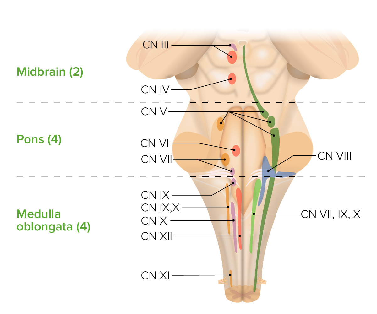
- Sympathetic ganglia
- Prosencephalon
- Spinal cord
Which area of the brain is responsible for arousal?
- Mesencephalon
- Telencephalon
- Metencephalon
- Myelencephalon
- Brainstem
A 5-year-old child is diagnosed with a medical condition that causes a deficiency in thyroid and growth hormones, and studies show developmental malformation in the hypothalamus. Which other structure might be affected?
- Thalamus
- Medulla
- Pons
- Corpus callosum
- Basal ganglia
A 72-year-old man had a stroke 3 months ago. Since then, he has experienced decreased motor ability and sensory perception on the right half of his body. What structure might have been injured?
- Left hemisphere
- Cervical spinal cord injury
- Right hemisphere
- Brainstem
- Corpus callosum
Researchers observe one particular area of the brain with increased activity when subjects read a book, describe intense emotional moments, or learn a foreign language. What is this area?
- Temporal lobe
- Right frontal lobe
- Cerebellum
- Left occipital lobe
- Parietal lobe
An autopsy of a patient's brain shows a growth in the right parietal lobe. How would the patient have been affected antemortem?
- Altered perception of sensory information
- Difficulty with speech
- Difficulty with comprehension
- Decreased vision in the upper quadrant of the visual field
- Altered emotional comprehension and planning
What change in neurotransmitters might be observed in a patient with depression?
- Increased acetylcholine
- Increased norepinephrine
- Increased serotonin
- Increased glutamate
- Increased dopamine
What neurochemical changes would be observed in a patient with panic attacks and anxiety?
- Decreased serotonin
- Decreased norepinephrine
- Decreased dopamine
- Decreased acetylcholine
- Increased acetylcholine
Author of lecture Structure and Function of the Central Nervous System – Biological Bases of Behavior (PSY, BIO)

Tarry Ahuja, PhD
Customer reviews
5,0 of 5 stars
| 5 Stars |
|
5 |
| 4 Stars |
|
0 |
| 3 Stars |
|
0 |
| 2 Stars |
|
0 |
| 1 Star |
|
0 |
