Playlist
Show Playlist
Hide Playlist
Secondary Lymphoid Tissues – Lymphocyte Activation
-
Slides Lymphocyte Activation.pdf
-
Download Lecture Overview
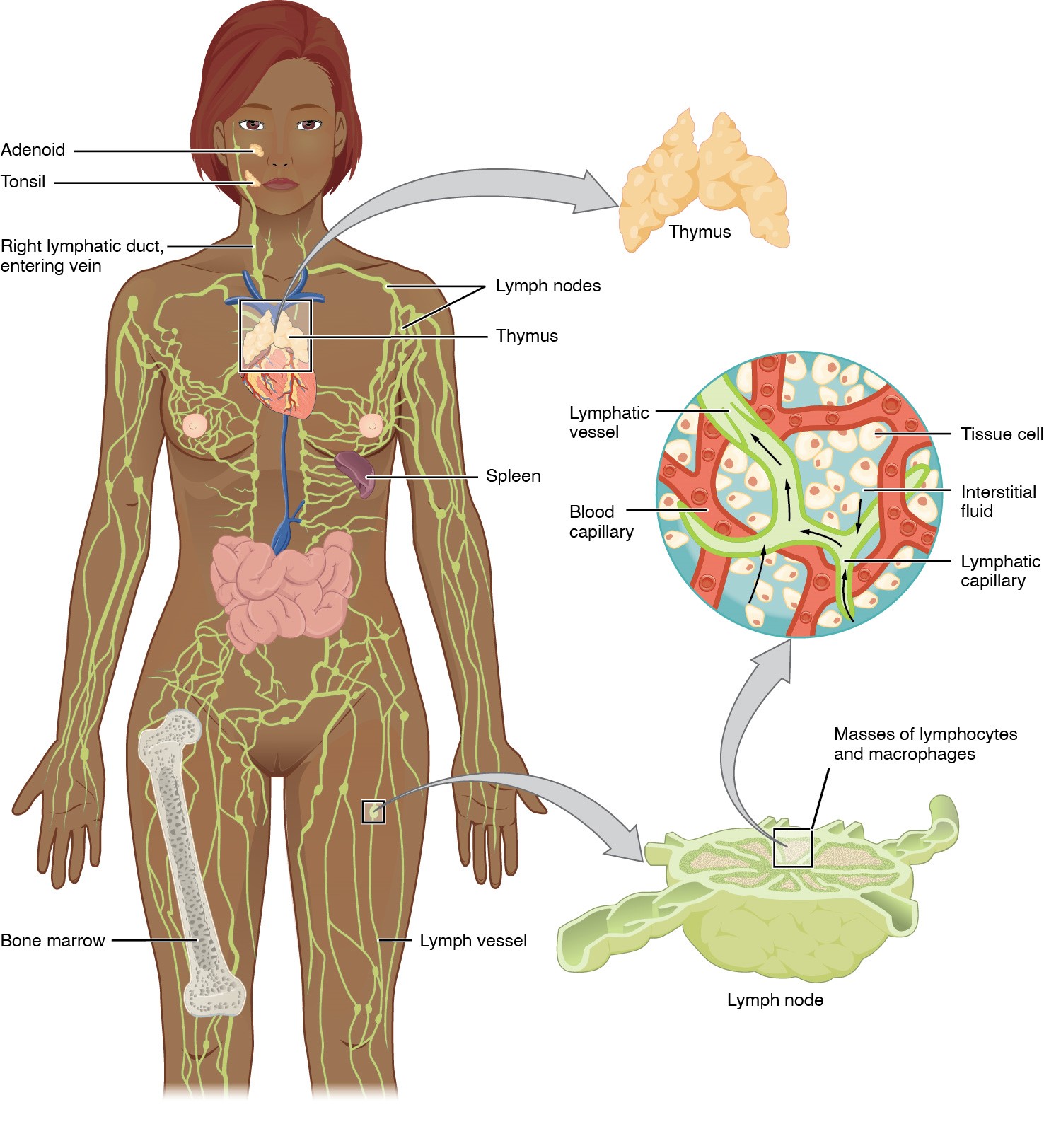
00:01 The secondary lymphoid tissues have structures that support the activation of the adaptive immune response. 00:12 There are a whole variety of cells within the secondary lymphoid tissues. 00:19 Different types of T-cells, B-cells, the antibody secreting plasma cells, dendritic cells which show antigen to T-cells, follicular dendritic cells which show antigen to B-cells, macrophages and natural killer cells; all of these cell types are present. So really it’s the secondary lymphoid tissues where the adaptive response becomes activated. So let’s look at these different types of secondary lymphoid tissue. Lymph nodes are scattered throughout the body. Lymphocytes can circulate around the body in the blood circulation and in the lymphatic circulation. And these two different circulations are not isolated from each other, there are connections between them. Particularly important being the thoracic duct which permits lymphocytes that are circulating around the lymphatic system to re-enter the blood circulation. Lymph nodes throughout the body are connected with one another via the lymphatic circulation, and they also all have a blood supply. So antigen can enter the lymph node either through the lymphatic vessels or potentially through the blood vessels. And likewise, lymphocytes and other cells of the immune response can enter the lymph node either via the lymphatics or via the blood circulation. The outside of the lymph node is surrounded by a collagenous capsule. Trabeculae penetrate into the inside of the lymph node from this capsule. Underlying the capsule is the subcapsular sinus. The inner part of the lymph node is referred to as the medulla. And there is a sinus where cells from the medulla and fluid from the medulla can drain into, and this is the medullary sinus. 02:36 Antigen, particularly antigen being carried by dendritic cells, can arrive in the lymph node via the afferent lymphatic vessel. 02:49 Lymphocytes can enter via the blood circulation, although they can also enter through the afferent lymphatics. 03:05 In order to enter the cellular part of the lymph node, the lymphocytes arriving via the arteries will enter through specialized structures referred to as high endothelial venules or HEV. 03:28 Once lymphocytes become activated within the lymph node, they organize themselves into follicles. 03:36 And particularly the B- lymphocytes produce follicles within the outer part of the lymph node, the cortex. 03:46 So the cortex constitutes the B-cell zone of the lymph node. 03:50 Within the cortex, the B-cells will eventually form into structures called germinal centers that develop in response to antigenic stimulation. 04:05 Cells can then exit from the lymph node following their activation, by using the efferent lymphatic vessels. 04:16 Turning now to the spleen; the spleen is comprised of red pulp, which contains erythrocytes and white pulp which contains the white blood cells of the immune system. 04:31 Trabecular arteries will feed into the white pulp. 04:37 There’s a central arteriole and a follicular arteriole by which lymphocytes can enter into the white pulp from the blood circulation. 04:52 The outer area of the white pulp is referred to as the marginal zone. 05:00 And there are marginal sinuses from which cells and molecules can drain. 05:08 The T-cell zone is referred to as the periarteriolar lymphoid sheath or PALS, P-A-L-S. 05:20 And the B-cells, again organize themselves into follicles, just like we saw in the lymph node. 05:27 Here we can see a tissue section of spleen and we can clearly identify the periarteriolar lymphoid sheath, a sheath of lymphocytes surrounding the artery. 05:42 Following antigenic stimulation just like in the lymph node, germinal centers can be produced. 05:50 Here in this immunofluorescent stained section, we can see the T-cell zone; here an antibody against the T-cell surface molecule CD3, an antibody coupled with a red fluorescent dye is identifying where the T-cells predominantly are located within the periarteriolar lymphoid sheath. 06:16 Whereas here, using an antibody against the cell surface molecule on the surface of B-lymphocytes, the CD20 molecule, you can see that there is a clear B-cell zone with a lymphoid follicle. 06:36 And finally, the Mucosa-Associated Lymphoid Tissues. 06:40 The MALT provides immunological protection of mucosal surfaces. 06:46 It involves lymphocytes and other immune system cells, and it's located at mucosal sites throughout the body. 06:57 For example, the GALT or Gut-Associated Lymphoid Tissue, which includes structures that are called Peyer's patches, the BALT or Bronchus-Associated mucosal tissue, which is usually only induced after infection. 07:16 And the NALT or Nasal-Associated mucosal tissue, as well as other types of mucosa associated lymphoid tissues. 07:26 Interspersed amongst the gut mucosal epithelial cells are specialised areas of lymphoid tissue referred to as Peyer’s patches. 07:35 These are closely associated with M cells, also referred to as microfold cells, which sample antigens in the gut and pass them to the underlying dendritic cells and macrophages. 07:48 The lymphoid follicles become more developed following activation of the T and B lymphocytes that are also present in the Peyer’s patches. 08:00 Once initially activated in this local environment, the lymphocyte will migrate to the nearest draining lymph nodes, which are the mesenteric lymph nodes.
About the Lecture
The lecture Secondary Lymphoid Tissues – Lymphocyte Activation by Peter Delves, PhD is from the course Adaptive Immune System. It contains the following chapters:
- The Secondary Lymphoid Tissues
- A Closer Look on the Lymph Nodes
- A Closer Look on the Spleen
- Mucosa-Associated Lymphoid Tissue
Included Quiz Questions
Where are the B cells mainly found in a lymph node?
- Cortex
- Paracortex
- Medulla
- Medullary sinus
- Subcapsular sinus
Which of the following is the main site of activation of the adaptive immune response?
- Secondary lymphoid tissue
- Blood
- Thymus
- Bone Marrow
- Mucosal surfaces
Which of the following types of vessels are found at the hilum of a lymph node?
- Artery, vein, efferent lymphatic
- Artery, vein, afferent lymphatic
- Efferent and afferent lymphatic
- Artery, vein, efferent lymphatic, and afferent lymphatic
- Artery and vein
Which of the following regarding the site of B cells and T cells within the spleen is CORRECT?
- The periarteriolar lymphoid sheaths (PALS) contain T cells and lymphoid follicles contain B cells.
- The periarteriolar lymphoid sheaths (PALS) contain B cells and lymphoid follicles contain T cells.
- The periarteriolar lymphoid sheaths (PALS) contain both B and T cells.
- The lymphoid follicles contain both B and T cells.
Customer reviews
5,0 of 5 stars
| 5 Stars |
|
5 |
| 4 Stars |
|
0 |
| 3 Stars |
|
0 |
| 2 Stars |
|
0 |
| 1 Star |
|
0 |


