Renal Clinical Anatomy
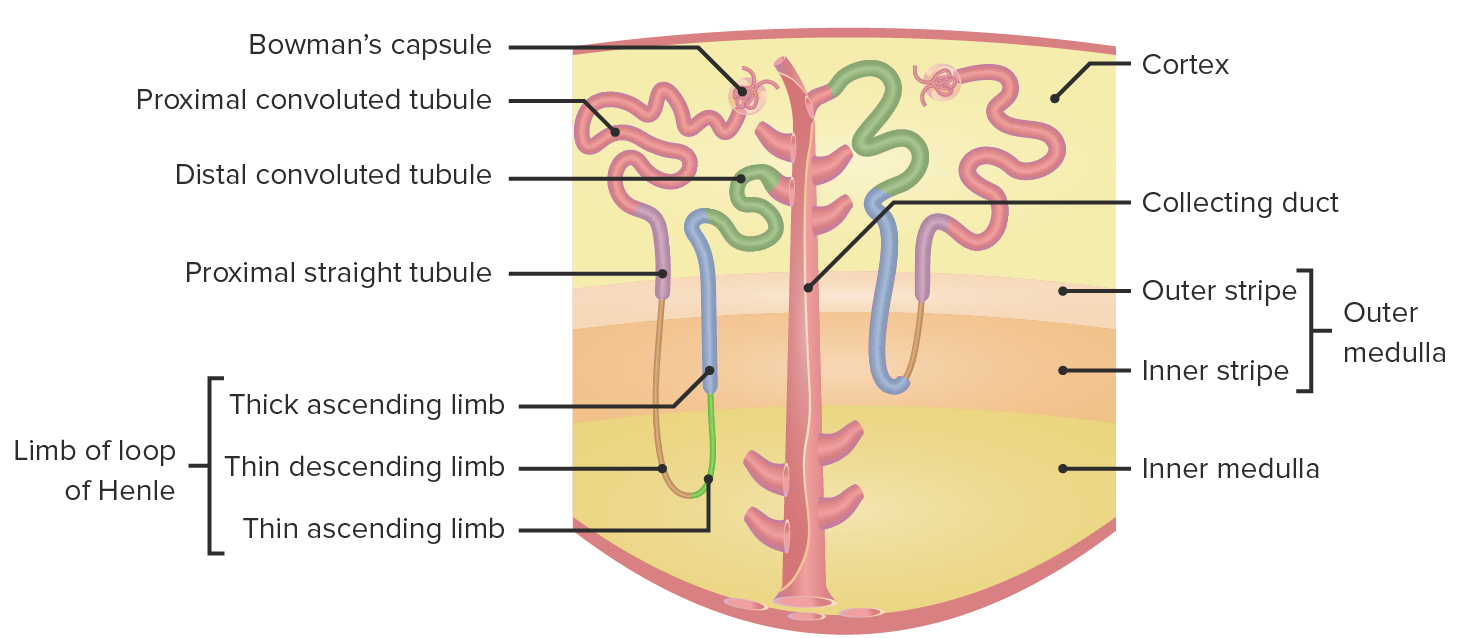
by Carlo Raj, MDA single kidney contains over one million functioning units called nephrons. A nephron consists of a glomerulus, which filters the blood free of cells and large proteins, and a tubule, a specialized series of ducts that act via absorption and secretion to produce urine from the glomerular ultrafiltrate. Homeostatic balance of electrolytes, acids, bases, and minerals is achieved via this complex balance of secretion, filtration, and reabsorption. Importantly, this process also rids the body of toxic waste products. Renal hormonal functions (e.g., calcium regulation, blood pressure regulation, and the production of red blood cells) are largely guided by cells outside of the nephron. Understanding renal clinical anatomy is paramount to understanding pathology.
Course Details
- Videos 4
- Duration 0:25 h
- Quiz questions 19
- Concept Pages 2