Playlist
Show Playlist
Hide Playlist
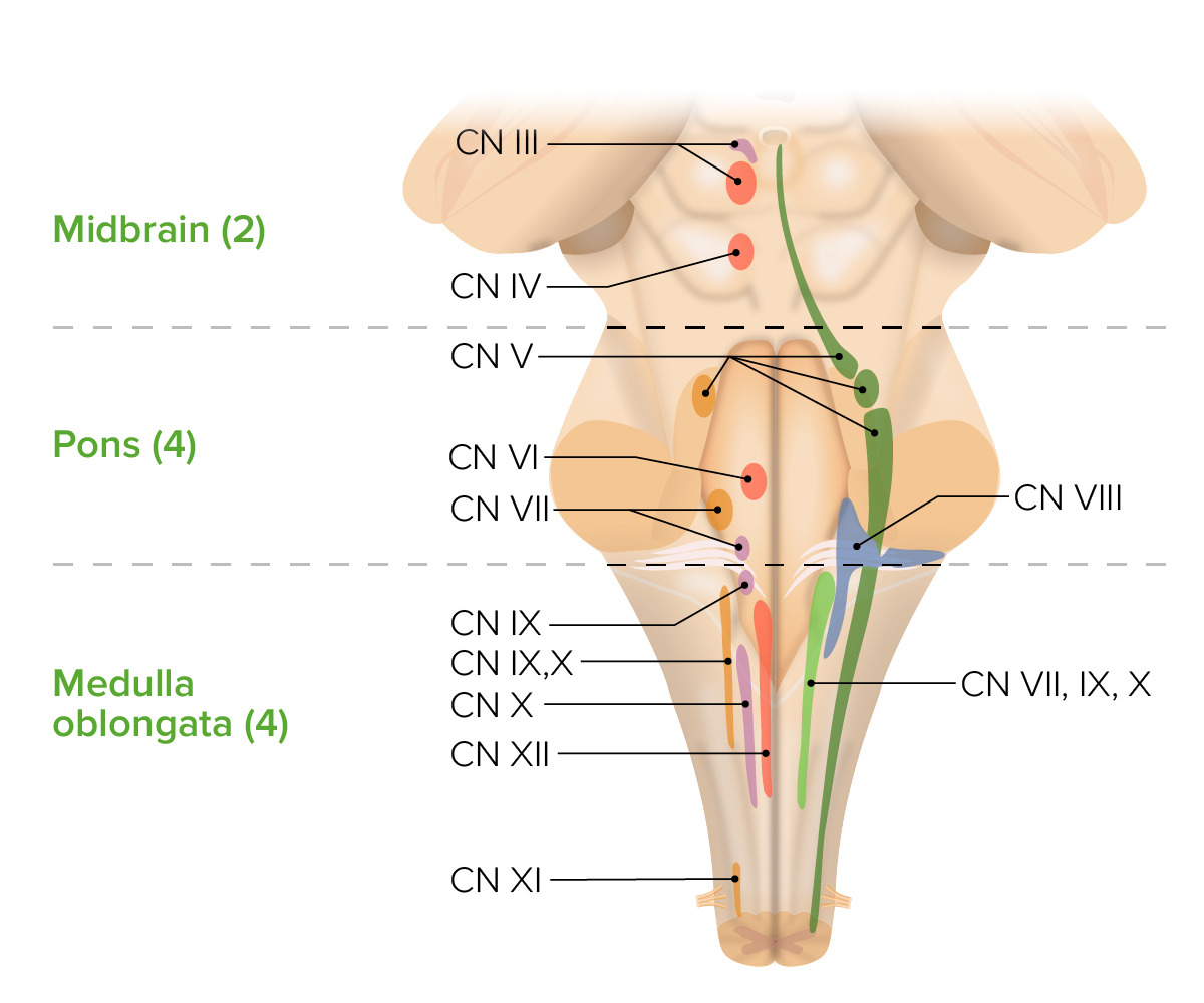
Posterior View of the Brain Stem

-
Slides 6 BrainStem BrainAndNervousSystem.pdf
-
Download Lecture Overview
00:00 Now, I want you to understand the posterior features of the brainstem. This is a posterior view that you are looking at here. We’re going to start in the midbrain area up in through here and then work our way down inferiorly. First structure that’s identified here for you would be the inferior colliculi. These are paired structures. These are important in orienting the head and ears to auditory stimuli. If you hear a sound coming from the right ear, you can orient your head and ears toward that incoming auditory stimulus. 00:46 You will move toward the right. The superior colliculus is also involved in orienting the head to a stimulus. 00:57 In this case, it’s going to be a visual stimulus. In addition to orienting the head toward the visual stimulus, the eyes will also move in that direction. If you kept something moving out of the left corner of your eye, your head and your eyes will move toward that moving object that you see off toward the left. 01:18 The trochlear nerve, cranial nerve number IV shown in through here. This was mentioned earlier. 01:26 This innervates the superior oblique muscle. This is the only nerve that issues from the posterior surface of the brain. Here we can find the pineal gland; it plays a role in the circadian rhythm. The circadian rhythm controls the 24 hour oscillation clock of behavioral, physical and mental changes associated with our sleep-wake-patterns. 01:57 Working our way down inferiorly, here we have a connection between the brainstem and the cerebellum. This is the superior cerebellar peduncle that we see in through here. This is going to provide the principal motor output of the cerebellum. This motor output will control or coordinate ipsilateral arm and leg movements. The middle cerebellar peduncle is shown here. This is the largest of the three peduncles. This is primarily conveying afferents from the pontine nuclei. Your last cerebellar peduncle is the inferior one. This is right in through here. There’s a little ridge here that separates the middle from the inferior. This is conveying afferents from the medulla and then also conveying efferents to the vestibular nuclei of the vestibular apparatus and pathway. Again continuing distally, here we can also visualize some additional structures. Here you’re looking at the floor of the 4th ventricle. 03:12 Here we have cuneatus structures. Here we have the fasciculus cuneatus. Then just a little bit superior to that, the tuberculum cuneatus. This is conveying fine touch, two-point discrimination, pressure, vibration, and conscious proprioception from the ipsilateral upper extremity. More medially located, we have gracilis structures. Here’s the fasciculus gracilis. Then a little higher here superior to that, we have the tuberculum gracilis. These gracilis structures are conveying fine touch, two-point discrimination, pressure, vibration, as well as conscious proprioception from the ipsilateral lower extremity.
About the Lecture
The lecture Posterior View of the Brain Stem by Craig Canby, PhD is from the course Brain Stem.
Included Quiz Questions
Which of the following cranial nerves arrises from the posterior aspect of the brain stem?
- Trochlear nerve
- Glossopharyngeal nerve
- Facial nerve
- Trigeminal nerve
- Abducens nerve
Which of the following structures, if damaged, could result in failure in orienting the head toward an auditory stimulus?
- Inferior colliculus
- Hippocampal gyrus
- Red nucleus
- Inferior cerebellar peduncle
- Superior cerebellar peduncle
Which of the following statements regarding cerebellar peduncles is most accurate?
- The inferior cerebellar peduncle receives afferents from the medulla and sends efferents to the vestibular nuclei.
- The superior cerebellar peduncle provides the motor output of the cerebellum to coordinate the contralateral arm and leg.
- The superior cerebellar peduncle is the only communication between the cerebellum and the brain stem.
- The middle cerebellar peduncle conveys efferents to the pontine nuclei.
- The inferior cerebellar peduncle is the largest of 3 peduncles.
Author of lecture Posterior View of the Brain Stem

Craig Canby, PhD
Customer reviews
5,0 of 5 stars
| 5 Stars |
|
5 |
| 4 Stars |
|
0 |
| 3 Stars |
|
0 |
| 2 Stars |
|
0 |
| 1 Star |
|
0 |
