Playlist
Show Playlist
Hide Playlist
Pediatric Appendicitis


-
Slides GD Pediatric GI.pdf
-
Download Lecture Overview
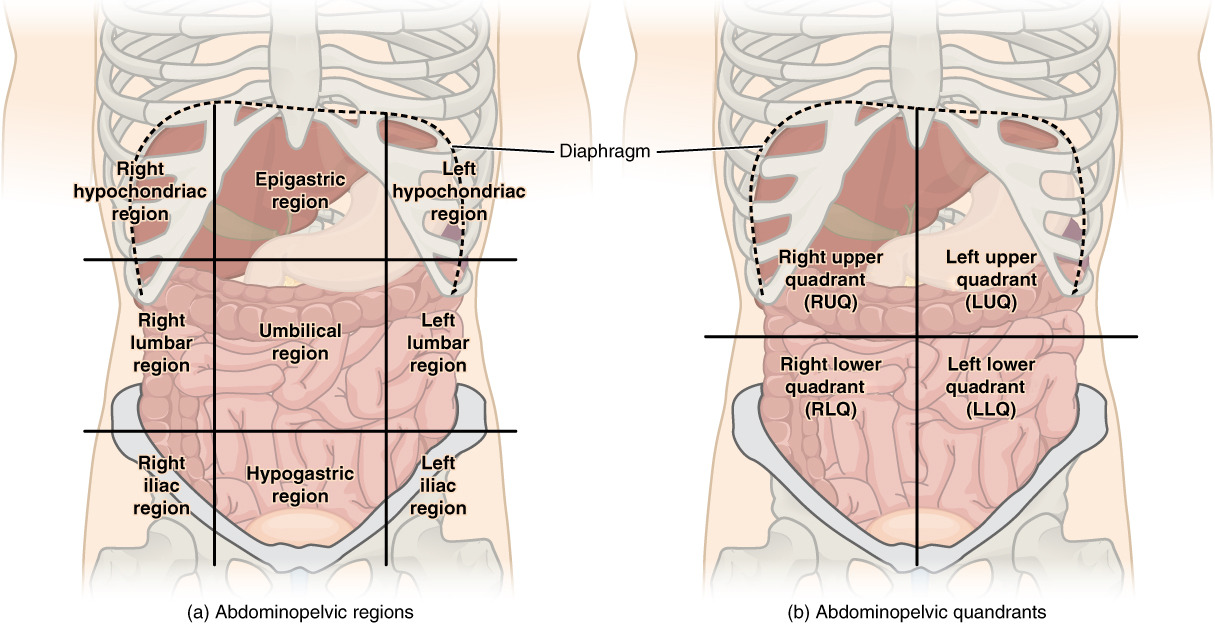
00:01 Our topic is pediatric appendicitis, not adult. 00:05 Pediatric. 00:07 Most commonly caused by lymphoid hyperplasia or perhaps fecalith. 00:12 What does that mean? Think about the appendix and know where you are. 00:16 Right lower quadrant. 00:18 What is an appendix? It’s vestigial “organ” and that at some point in time, it behaves exactly like a diverticulum. 00:29 And so therefore, if you have feces passing through could it get stuck in the appendix? Sure, it can. 00:35 Welcome to fecalith. 00:37 And highlight or keep in your head, lymphoid hyperplasia being a major cause of appendicitis in this child. 00:46 It may then cause abdominal pain. 00:50 Here, signs and symptoms, the child with this pain is going to vomit. 00:56 Emesis, fever, right lower quadrant pain, peritoneal signs after 36 hours, what does that mean to you? Oh, my goodness, there’s every possibility that the appendicitis is going to rupture. 01:12 With this type of rupture with an appendicitis, what is it going to do? You’re going to have a massive WBC reaction. 01:21 What is this reaction called, please? Good. 01:24 Leukemoid reaction. 01:27 And your WBC count, why do we call this leukemoid? It looks like leukemia. 01:34 Your WBC count could be as high as 50,000. 01:39 What’s your normal WBC count? 4,500 to 11,000. 01:44 Do not forget about peritoneal sign. 01:47 Diagnosis, look at some differentials here. 01:49 Remember, a yersinia infection -- do not forget this -- a yersinia infection, a gastroenteritis that behaves like appendicitis. 01:59 Remember, pseudo-appendicitis with yersinia and inflammatory bowel disease is a possible differential. 02:07 Keep these in mind with differential diagnoses. 02:14 Diagnosis itself, CBC, appendicitis, you find it be elevated. 02:19 Upon ultrasound, what you’re seeing here is thickening of the appendix, that’s what the arrows are pointing to. 02:26 Whereas the CT here, remember where you are remember how to interpret a CT? The left side of this image is the right side of the patient. 02:40 Your appendix located in the right lower quadrant. 02:43 You’ll notice the white area there which has become inflamed and that is going to be your appendicitis on CT. 02:52 Ultrasound, CT for proper diagnosis. 02:57 Management: Always worry about infection. 03:00 Antibiotics IV. 03:02 Surgery, you need to get in there and remove this before this thing ruptures. 03:07 Pediatric appendicitis, lymphoid hyperplasia.
About the Lecture
The lecture Pediatric Appendicitis by Carlo Raj, MD is from the course Pediatric GI Pathology.
Included Quiz Questions
A 10-year-old boy comes to the emergency department because of a 2-day history of severe right lower quadrant pain, fever, nausea, and vomiting. He lives in the woods, and his parents do not have direct access to health care facilities. He collapses on reaching the hospital. On blood testing, which of the following would you expect?
- Leukemoid reaction
- Lymphoid reaction
- Myeloid leukemia
- Lymphoid leukemia
- Monocytosis
Which of the following is NOT a common complication of appendicitis?
- Intussusception
- Rupture of appendix
- Gangrene of appendix
- Appendicular mass
- Peritonitis
Author of lecture Pediatric Appendicitis

Carlo Raj, MD
Customer reviews
5,0 of 5 stars
| 5 Stars |
|
5 |
| 4 Stars |
|
0 |
| 3 Stars |
|
0 |
| 2 Stars |
|
0 |
| 1 Star |
|
0 |
