Playlist
Show Playlist
Hide Playlist
Neurofibromatosis, Hypotonia and Childhood Neurodegenerative Disorders

-
Slides 13 PediatricNeuropathology Neuropathology II.pdf
-
Download Lecture Overview
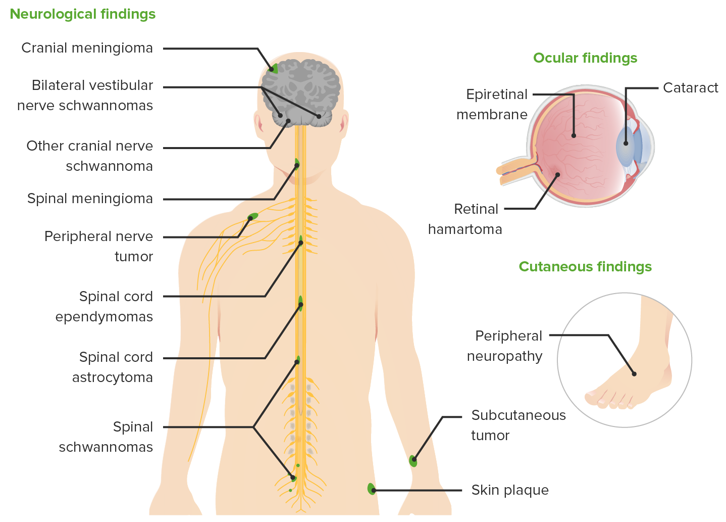
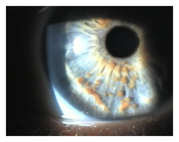
00:00 So neurofibromatosis type 1. When we did our primary brain tumors, at some point in time we looked at schwannoma. Schwannoma was arising from a neurofibromatosis type 2 and at that time with that type 2, I told you to please memorize a particular gene that it quotes for called merlin. Here, we have a neurofibromatosis type 1. The name of the gene is NF1 neurofibromin. 00:35 Incidence 1 in 3000 and the name of the gene or protein is called neurofibromin, number 1 neurofibromin. Merlin type 2. Those are important. With neurofibromatosis type 1, our topic here is neurocutaneous, we're talking about children or young pediatric neuropathology as the topic. Isn't it? These mocha-colored macules that you might find on the skin, mocha colored. What color is that? Darken mocha, cafe au lait spots, axillary freckles, Lisch nodules, or iris hamartomas. These are important specifically for neurofibromatosis type 1. Another name for neurofibromatosis type 1 is called von Recklinghausen, plexiform and simple neurofibromas. What does that mean? Neurofibromas are benign nerve sheath tumors that can be found in patients with neurofibromatosis type 1. They are different from schwannomas in that they incorporate many additional types of cells and structural elements in addition to Schwann cells. You can have different types of neurofibromas. These may include, I'm giving you a couple, plexiform and simple. But really the point is an osis, fibromatosis, goes on to neurofibroma, optic glioma, obstructive type of hydrocephalus, and pheochromocytoma could be all part of the syndrome. In neurofibromatosis type 1, once again please don’t forget about the protein, it's called neurofibromin. Where is neurofibromatosis type 2 which we've talked about already? Also, autosomal dominant? Much less common than NF1 but you must know it. 02:33 Allow the 2 to speak to you and by that I mean type 2, 2 ears schwannoma, acoustic neuroma. 02:41 The name of that protein here is called schwannomin or merlin. Type 2, the chromosome is 22 merlin. Memorize that please. With type 2, if it goes on to cancer, what is it? There you go, bilateral acoustic neuroma aka schwannoma. Multiple meningiomas could also be a possibility. 03:08 Keep that in mind please. Genetic testing available for both types of NF neurofibromatoses and also available for tuberous sclerosis complex. What does that mean to you? At least no hamartin. And I told you about the skin issues and tuberous and so on and so forth with TSC. 03:30 Our topic here is hypotonia, reduction in pustule in other words a floppy baby. May not be associated with significant weakness by definition. It could be central or peripheral in terms of cause where you find hypotonia in a child. Our topic is pediatric neuropathology. Under hypotonia, if it's central in a child you're usually looking for associative signs of CNS dysfunction; seizures, developmental delay. What does that mean to you? You're not reaching a particular milestone, you keep to separate from intellectual disability formally known as mental retardation, microcephaly and dysmorphic features. If it's peripheral hypotonia, lack of central signs, weakness is usually prominent. Prominent here with peripheral. Here you might be thinking about spinal muscular atrophy, SMA; peripheral neuropathy; neuromuscular junction abnormalities, or perhaps even myopathies. This is the topic, hypotonia in pediatric population. The childhood neurodegenerative disease is one that you want to take a look at and have differentials. If you notice macular cherry red spots, you're thinking about your hexosaminidase deficiency, welcome to Tay Sachs disease, if it's single myelin and it's Niemann-Pick disease. If you find glucocerebrosidase deficiency, the most common lysosomal storage disease, the patient could have Gaucher disease or metachromatic leukodystrophy. 05:02 Could be found, well macular cherry red spot, would do a fundoscopic examination and you find a red spot in the retina, you're a differentials. Are we clear?
About the Lecture
The lecture Neurofibromatosis, Hypotonia and Childhood Neurodegenerative Disorders by Carlo Raj, MD is from the course Pediatric Neuropathology. It contains the following chapters:
- Neurofibromatosis Type 1
- Neurofibromatosis Type 2
- Hypotonia
- Childhood Neurodegenerative Disorders
Included Quiz Questions
Neurofibromatosis type 2 is caused by a defect in the gene that normally gives rise to which of the following proteins?
- Merlin
- Fibrillin
- Neurofibromin
- Tuberin
- Hamartin
Which of the following cranial nerves is particularly involved in neurofibromatosis type 2?
- Vestibulocochlear
- Olfactory
- Abducens
- Trigeminal
- Facial
Author of lecture Neurofibromatosis, Hypotonia and Childhood Neurodegenerative Disorders

Carlo Raj, MD
Customer reviews
3,0 of 5 stars
| 5 Stars |
|
0 |
| 4 Stars |
|
0 |
| 3 Stars |
|
1 |
| 2 Stars |
|
0 |
| 1 Star |
|
0 |
The speech style is a little robotic? Maybe this would be better as an audio book.
