Playlist
Show Playlist
Hide Playlist
Lymphadenopathy: Multiple Myeloma (Introduction)


-
Slides Lymph Nodes.pdf
-
Download Lecture Overview
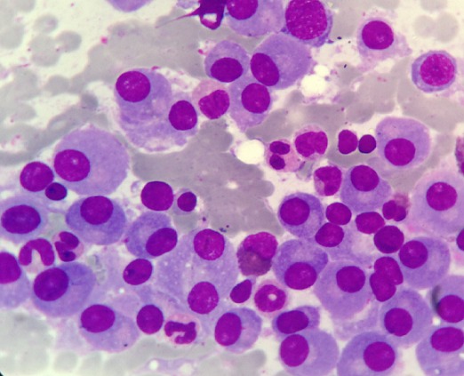
00:01 A plasma cell neoplasia that is known as multiple myeloma is what we have here. 00:05 Remember your clinical presentation, you are definitely going to have a patient that comes in with lytic bone lesions, is gonna have those osteolytic lesions, will have pathologic fractures and because of that will result in hypercalcemia. 00:18 The products secreted include the following. 00:22 You have an intact monoclonal immunoglobulin, now there’s something called “M protein,’’ the M protein not to be confused with IgM please. 00:33 The M protein would be the combination of the fact that you’re forming your light chains, kappa and lambda, if this pair of proteins and such all these along with the calcium will then give rise to what we call in multiple myeloma as M protein. 00:47 The type of immunoglobulins that you produce with multiple myeloma no doubt is IgG or IgA it’s not IgM, I repeat once again, it is not IgM. 00:58 Next, you’re not going to have heavy chains here that you’re going to dump into the urine that the fact that you have light chains, these light chains include kappa and lambda and 70% of these would be significantly, please no kappa, kappa, kappa - these free light chains which would then deposit on, let’s say you glomerulus or will then filtering get into the urine we then call Bence-Jones proteinuria as you're quite familiar with. 01:28 And of course that light chain, those light chains that accumulate on the glomeruli, you would then do a congo red stain and then you do immunofluorescence and lo and behold what do you find? Apple green birefringence because these amyloid, right, apple green birefringence. 01:43 Now, what you’re looking at here on the right is your serum protein electrophoresis. 01:48 What you’re seeing on top from the cathode, positive on the left over to the right anode is a normal protein that you can expect in our body. 01:58 These then include abdomen being the most abundant coming from the liver and nearby the protein such as alpha 1 antitrypsin, alpha 2 beta get from fibrinogen but most importantly for our understanding we have the gamma. 02:11 What you see there is a gamma wave, perfectly normal by the way and that gamma wave then represents five immunoglobulins. 02:18 These then do include IgG, IgA, IgM, IgE, IgD -- in other words, I’ve given you all immunoglobulin, G, A, M, E, D - but please be careful that gamma doesn’t represent only IgG, it represents all immunoglobulins, that is important. 02:35 Now, you can imagine in multiple myeloma that you have monoclonal gammopathy. 02:39 You are producing excess levels of IgG or IgA. 02:43 You’ll notice now at the bottom serum protein electrophoresis and lo and behold, take a look at the gamma wave and it is now spiked. 02:49 Somebody call this a gamma spike and that gamma spike of course refers to either IgG or IgA and does not represent IgM.
About the Lecture
The lecture Lymphadenopathy: Multiple Myeloma (Introduction) by Carlo Raj, MD is from the course Lymphadenopathy – White Blood Cell Pathology (WBC).
Included Quiz Questions
An abnormal kappa to lambda light chains ratio is a sensitive indicator for which of the following diseases?
- Multiple myeloma
- B-cell lymphoma
- Acute lymphoblastic leukemia
- Chronic myeloid leukemia
- Hodgkin lymphoma
Which of the following proteins is most abundant in human plasma?
- Albumin
- Fibrin
- Immunoglobulin M
- Globulin
- Heptoglobin
In serum protein electrophoresis, which protein is represented by the largest peak closest to the positive electrode?
- Albumin
- Gamma
- Alpha-1
- Beta-1
- Alpha-2
Which protein, when found in urine, may be suggestive of multiple myeloma?
- Bence-Jones protein
- Coronin
- Amyloid
- Albumin
- Globulins
Author of lecture Lymphadenopathy: Multiple Myeloma (Introduction)

Carlo Raj, MD
Customer reviews
5,0 of 5 stars
| 5 Stars |
|
5 |
| 4 Stars |
|
0 |
| 3 Stars |
|
0 |
| 2 Stars |
|
0 |
| 1 Star |
|
0 |
