Playlist
Show Playlist
Hide Playlist
Lesions of the Visual Pathway


-
Slides 15 VisualPathway BrainAndNervousSystem.pdf
-
Download Lecture Overview
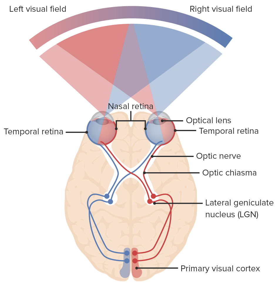
00:00 Now along this journey, light transmission can encounter some hazards. So there can be some lesions that will block this journey and result in various forms of visual impairments. So we’re going to take a look at various visual impairments based on the location of the lesion. So first, we’re going to have a lesion of the entire optic nerve. That lesion is shown right here. 00:37 In this case, the lesion will produce a loss of vision in the right eye, anopia. If we look here, this is the right eye, this is the left eye and we can see that the right eye has gone dark, black. 00:57 It’s not perceiving anything or it’s not conveying anything to the primary visual cortex. 01:03 Transmission’s been blocked completely here. What are some of the causes of this type of lesion? There are a couple. One could be trauma or another one could be inflammation of the optic nerve, so we have optic neuritis. Here, we’re going to shift the lesion to the left optic nerve. 01:30 It’s not going to be a complete lesion as we saw with the right optic nerve. Instead, it’s going to be a partial lesion of the left optic nerve. It’s going to be this lateral half. 01:43 So if we take a look at what’s going on here, here’s your temporal retina. This is the path that’s being blocked. So just the left temporal retina is being blocked in that pathway. What will happen here is you'll have a loss of the left temporal retina. That will result in blindness of the left nasal visual field. That is shown right in through here where this area of the left eye has gone dark. This is left nasal hemianopsia. Causes of this visual deficit could be a tumor just compressing the lateral side of the left optic nerve or it could be a form of arterial dysfunction and so there’s a disruption in the blood supply to just this lateral component of your optic nerve. 02:49 This could occur also on the right side. Another form of a lesion is where the lateral portion of the right optic nerve is damaged at the optic chiasma. That is shown right in through here. 03:13 So if we look at how the axons are travelling through this area, we will see that the nasal retina of the right eyeball right in through here, this pathway is uninterrupted. It crosses over here and then continues to the left primary visual field. What is disrupted, however, is the temporal retina from the right eye. See, that pathway is blocked. You’re also going to get a blockage of the nasal retina from the left eye. It’s crossing over here and then the lesion is sufficient enough to interrupt its transmission of the action potentials to the primary visual cortex. 04:08 What will happen here is you’ll have a loss of right temporal retina and blindness of the right nasal visual field. That is shown right in through here. This is right nasal hemianopsia which are also involving the left eye as well. Its deficit is this area here which is a left upper temporal quadrant anopsia. So this part goes black or dark as it can no longer perceive this visual information. This disorder is kind of like a slice of a pie. So this is referred to as pie in the sky disorder when we have this left upper temporal quadrant anopsia. 05:01 Cause of this: again, we’re looking at a structure that would lie lateral to the optic chiasm. 05:11 Our primary culprit here is our internal carotid artery. So if there’s a weakness in the wall of the internal carotid artery, there’s an aneurysm, you have dilatation. That aneurysm can put pressure on just this general area here and cause this kind of deficit. This could occur on the opposite side as well. Here, we have a simpler lesion in that the optic chiasm has been lesioned completely. We see that lesion right in through here. So any of the fibers that are crossing are blocked at this particular point. This will cause bitemporal hemianopsia. 05:59 So, you see the temporal regions here of the left and right eyes going dark. 06:09 Causes? Again, you’ve got to think about what’s in this area. You have the pituitary gland. 06:17 So an expansion of a pituitary tumor could put pressure on the optic chiasm and cause this type of lesion. Another type of tumor is a craniopharyngioma. That too can grow and involve the optic chiasm causing this lesion. Now, we’re looking at a lesion of the optic tract. 06:49 This tract runs from the optic chiasm to the lateral geniculate body on each side. So here’s the right one. This is the one that’s involved. So we see a complete lesion then through this right optic tract. This will cause left homonymous hemianopsia. What goes dark here would be the temporal retina of the left eye and then the nasal retina of the right eye. 07:29 Causes of this form of lesion would be a tumor of the temporal lobe because the temporal lobe is in this area. So if the tumor grows into this right optic tract, that could cause a lesion. 07:43 If there’s an abscess of the temporal lobe, that abscess can also disrupt the transmission of our visual pathway along the right optic tract. Of course, this can occur on the left side as well. Another point at which a lesion can occur is along the optic radiations. 08:11 The optic radiations can be completely lesioned or they may be partially lesioned. We’re going to look at a partial lesion, two types of partial lesions of our optic radiations. The first one here is a lesion of the right upper optic radiations. These are a part of Baum’s loop. They’re starting to terminate on your primary visual cortex in this area. So this could be a side of the lesion here or it could be a little more proximal or closer to the lateral geniculate bodies. This will result in left homonymous inferior quadrant anopsia. If we take a look here, here’s the quadrant that’s disrupted in the left nasal retina. Then here’s a quadrant that’s disrupted on the right temporal retina. Causes here could be a parietal lobe tumor or an occipital lobe tumor putting pressure on this part of the pathway. Lesions can also occur along the lower optic radiations as we see in through this area here. This is also starting to include the upper, so we kind of want to just look at this right in through here. Lesions of the lower optic radiations, these axons that are travelling here form Meyer’s loop. They will result in a darkness here and here of each eye. This is termed left homonymous superior quadrant anopsia. 10:18 Causes of this would be a tumor. It could be a tumor of the temporal lobe or a tumor of the occipital lobe. Then, of course, you can have a disruption of the entire optic radiations. 10:37 In here we have a complete lesion on the right side of the optic radiations. This then results in left homonymous hemianopsia. So you can see the darkness here in the left eye and then the disruption here at the right eye. Causes would be tumors, could have arterial dysfunction, interruption of blood flow to this area, or a trauma injury to the brain in this particular area. 11:18 What are some of the causes of this lesion? First, we can have tumors again disrupting this final pathway to the primary visual cortex. Arterial dysfunction could also cause a lesion to the entire optic radiations. Trauma is another possibility or cause to this particular visual deficit.
About the Lecture
The lecture Lesions of the Visual Pathway by Craig Canby, PhD is from the course Visual Pathways.
Included Quiz Questions
Which of the following visual field defects is caused by a partial lesion of the left optic nerve fibers?
- Blindness of the left nasal visual field
- Complete blindness of the left eye
- Blindness of the right nasal visual field
- Blindness of the right temporal visual field
- Complete blindness of the right eye
Which of the following visual field defects is caused by a median lesion at the level of the optic chiasm?
- Bitemporal hemianopsia
- Right temporal hemianopsia
- Binasal hemianopsia
- Right nasal hemianopsia
- Left temporal hemianopsia
Which of the following visual field defects is most likely caused by a tumor of the left temporal lobe compressing the left optic tract?
- Right homonymous hemianopsia
- Bitemporal hemianopsia
- Blindness of the left eye
- Left homonymous hemianopsia
- Binasal hemianopsia
Which of the following visual field defects is most likely caused by a tumor of the pituitary gland?
- Bitemporal hemianopsia
- Binasal hemianopsia
- Blindness in both eyes
- Right homonymous hemianopsia
- Left homonymous hemianopsia
Which of the following visual field defects is most likely caused by an occipital lobe tumor compressing the right lower optic radiations (Meyer's loop)?
- Left upper-quadrant anopsia
- Central scotoma
- Left homonymous hemianopsia
- Left lower quadrant anopsia
- Right homonymous hemianopsia
Author of lecture Lesions of the Visual Pathway

Craig Canby, PhD
Customer reviews
4,7 of 5 stars
| 5 Stars |
|
2 |
| 4 Stars |
|
1 |
| 3 Stars |
|
0 |
| 2 Stars |
|
0 |
| 1 Star |
|
0 |
Found this video very helpful. I finally understand how these lesions affect our vision. Thanks
After reviewing Neuroanatomy in FA and Kaplan, it is fair to say this lecture has delved deeply in some accurate key points of visual pathways.I really admire the fact that you guys have put on some quizzes in the end but I was surprised to see a red flag that goes unnoticed.In one of the question about the right lower optic radiations from the 5 listed, I saw right after radiation Meyer's loop in parenthesis which is clearly a mistake when it comes to asking questions.In the exam, they won't give any clues to test takers because they will have figure out themselves whether or not it is either Meyer's or Non-Meyers' loop.
Explains it to a point where the visual pathway is simple.
