Playlist
Show Playlist
Hide Playlist
Isovolumetric Contraction: LV Phases— Cardiac Cycle


-
Slides CardiacCycle CardiacPhysiology.pdf
-
Download Lecture Overview
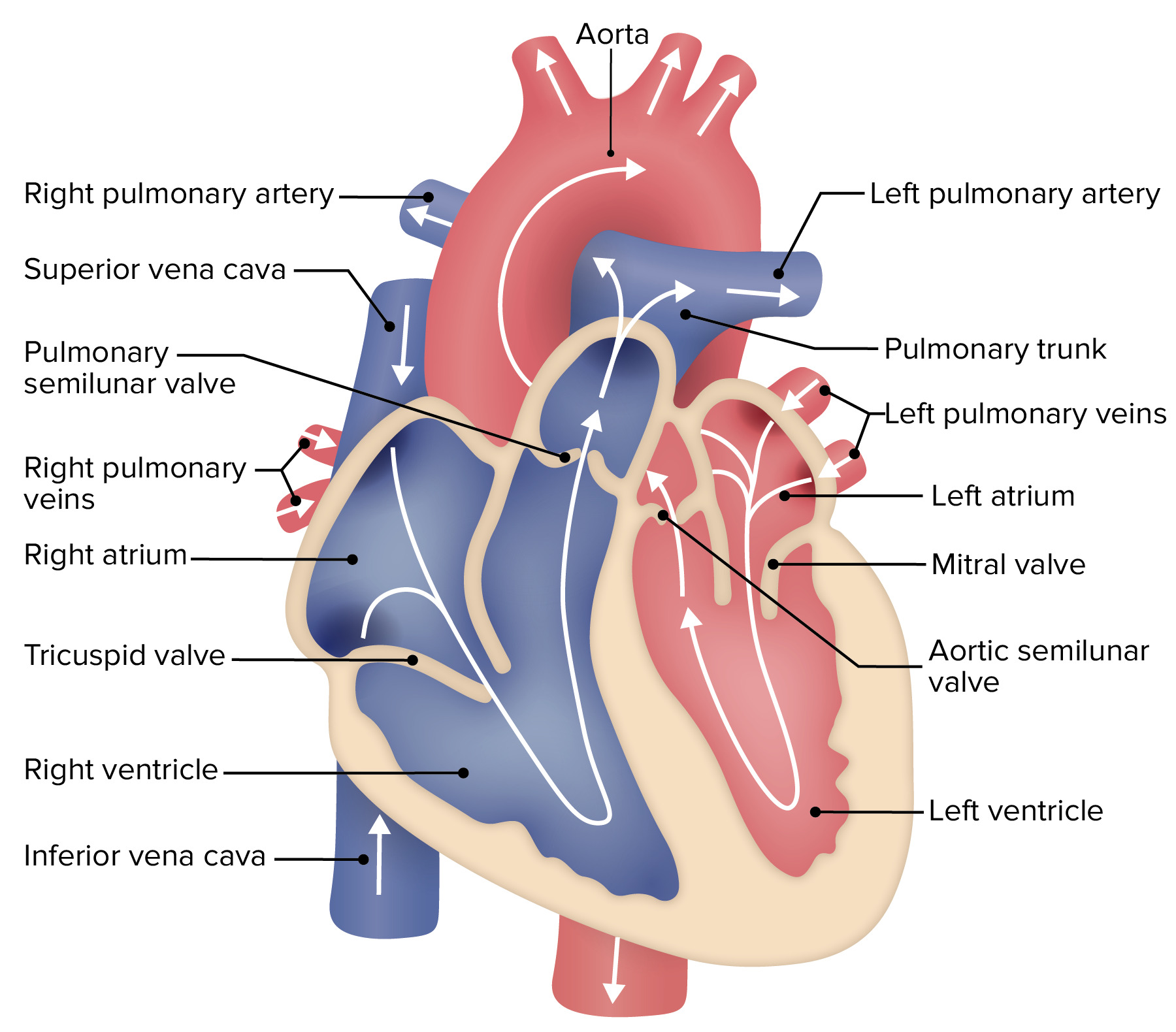
00:01 This is the classic cardiac cycle. 00:05 For this, we’re going to start off in isovolumic contraction. 00:10 So, I know these terms are a little bit difficult, but you must invest the time in to learn them because this is how everyone is going to refer to the various stages of the cardiac cycle and when blood is being pumped out. 00:26 What does isovolumic mean? Iso meaning the same, volumetric is the volume is the same during a contraction. 00:37 Isovolumetric contraction. 00:40 Thus, whatever is being filled, this is the left ventricular end-diastolic volume. 00:48 What we have is then a depolarization of the left ventricle. 00:52 And remember, that's the QRS complex from your ECG. 00:57 At this point, the left ventricular pressure increases, which is the pressure within the left ventricle. 01:05 The mitral valve closes, and this left ventricular volume then is maintained throughout this particular portion of the cycle. 01:16 The big thing that looks like that's changing here is that left ventricular pressure is increasing. 01:23 So, the volume doesn't change, just the pressure. 01:27 Now, we enter force of ejection. 01:31 There's going to be a rapid ejection, which is number three. 01:34 There's going be reduced ejection, which is number four. 01:37 When does ejection happen? As soon as the aortic valve opens. 01:42 So, you build up pressure within this left ventricle, you’re squeezing in on this fluid, but the fluid is not traveling anywhere and then, BOOM, pops through the aortic valve. 01:52 At that point, blood is now ejected into the aorta and then out through the systemic vasculature. 01:59 What happens here in terms of the ECG? This is at the time when the T wave occurs. 02:05 And remember from the ECG, the T wave is during ventricular repolarization. 02:12 The last item to kind of consider is that left ventricular end-systolic volume will be at the lowest point at the end of the reduced ejection or phase 4. 02:25 So, where do you get left ventricular end-diastolic volume? That is the spot that you started with for isovolumic contraction. 02:33 Where do you get left ventricular end-systolic volume? That’s the last point of the ejection phase. 02:45 Isovolumic relaxation is the next phase, and this is when the aortic valve closes. 02:52 So, once again, you're at a place where both valves are closed, both aortic and the mitral valve, and they will be closed until the pressure drops to a low enough level to open up that mitral valve. 03:06 Notice here that the volume again doesn't change, but the pressure is rapidly dropping. 03:13 The next phases are the ventricular filling. 03:17 Ventricular filling is so important. 03:19 We almost always overlook it. 03:21 Why? Because you need to fill up the left ventricle before you have anything to contract. 03:27 Therefore, that's the bolus of blood that has returned to the left ventricle. 03:31 We break it up into a couple of different components. 03:34 One is a rapid filling and the other is a reduced filling. 03:40 That is, six is rapid, seven is reduced. 03:44 You notice here that it's a longer phase. 03:47 That's because the heart spends more time in diastole than it does in systole. 03:52 So, if you want to think about it in terms of the speed, think of this. 03:56 Systole. 03:57 D-i-a-stole. 03:59 Systole. 04:00 D-i-a-stole. 04:03 The filling phase always accounts for more time than the contraction phase. 04:09 Hopefully, you can appreciate through this type of graph that there is a large amount of volume going into that left ventricle. 04:19 Interestingly, if you haven't thought about this before, if you think about the filling of the left ventricle, most of the time, when you first learn this, you always feel that, okay, it fills a little bit and then the top part of the heart contracts and pushes in the rest of the blood, right? Well, not so much. 04:40 The ventricular filling component encompasses most of the diastolic filling, probably 90% of it is done passively. 04:49 You only add about 10% through atrial contraction. 04:55 And that is shown here, the atrial contraction. 04:58 You see a very small blip in pressure and that increases about the last 10% of ventricular filling. 05:08 When we’re trying to quantify how much blood leaves the heart, it's probably best to do that in something called an ejection fraction. 05:16 An ejection fraction, this formula, is just a concept of how this process works. 05:23 It's usually measured with echocardiography. 05:27 So, you can put someone and measure their left ventricular end-diastolic volume, the left ventricular end-systolic volume, you divide those – that quantity by end-diastolic volume again. 05:38 And that gives you a fraction. 05:41 And that fraction should be, in the average person, somewhere between 55 and 60. 05:47 However, if you have heart failure, that is a person that has a low ejection fraction, and this can be very dramatic or it can just be a partial decrease in ejection fractions. 06:00 So, ejection fraction is an important clinical monitor for looking at the volume that leaves the left ventricle.
About the Lecture
The lecture Isovolumetric Contraction: LV Phases— Cardiac Cycle by Thad Wilson, PhD is from the course Cardiac Physiology. It contains the following chapters:
- LV Systole
- LV Diastole
Included Quiz Questions
Which of the following stages of the cardiac cycle does the ventricular volume curve drop sharply?
- Ejection
- Atrial contraction
- Isovolumic contraction
- Passive filling
- Isovolumic relaxation
Where do you measure left ventricular end-diastolic volume and left ventricular end-systolic volume, respectively?
- Isovolumic contraction and the last point of the ejection phase
- Isovolumic relaxation and mid-ejection phase
- Mid-filling phase and the last point of the ejection phase
- The last point of the ejection phase and isovolumic relaxation
- Both can be measured in the filling phase.
What is the normal range of the ejection fraction?
- 55-60%
- 15-25%
- 25-35%
- 80-90%
- 30-40%
Author of lecture Isovolumetric Contraction: LV Phases— Cardiac Cycle

Thad Wilson, PhD
Customer reviews
5,0 of 5 stars
| 5 Stars |
|
1 |
| 4 Stars |
|
0 |
| 3 Stars |
|
0 |
| 2 Stars |
|
0 |
| 1 Star |
|
0 |
great lecture realy useful im first year dentistry school in romania
