Playlist
Show Playlist
Hide Playlist
Hydrocephalus: Definition and Treatment


-
Slides 2 VentricularSystem BrainAndNervousSystem.pdf
-
Download Lecture Overview
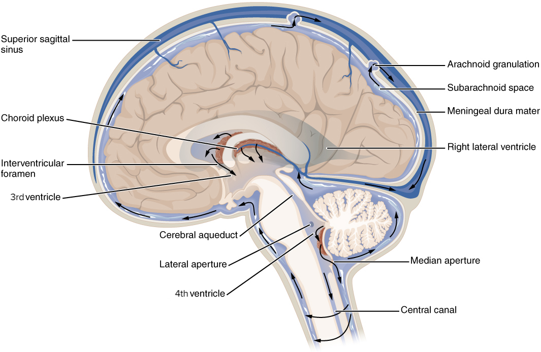
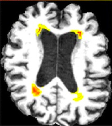
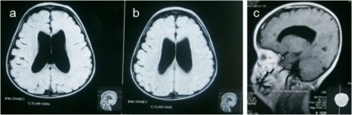
00:00 The image that you see to your left is showing a normal amount of cerebrospinal fluid. This is an axial section of the brain. You can clearly see portions of the lateral ventricles. This would be an anterior view. 00:18 This would be a posterior view. If there’s an excess in cerebrospinal fluid, there will be a dramatic increase in cerebrospinal fluid such that the lateral ventricles in this view are going to become greatly dilated or distended. 00:35 So this is a case of hydrocephalus. You can see how dilated these lateral ventricles are compared to a normal state. 00:47 Hydrocephalus is due to a disturbance of formation, flow, absorption of cerebrospinal fluid. Some causes of this disruption would be first, obstruction of the cerebral aqueduct. So in this case, cerebrospinal fluid is constantly being produced in the lateral ventricles but it cannot move from the third ventricle into the fourth. So some of it can pass into the third but that’s as far as it gets. Then the lateral ventricles will become greatly distended as we see here. Arnold–Chiari malformation can also disrupt cerebrospinal fluid flow so that it excessively accumulates. 01:39 Dandy-Walker malformation is another example that can cause hydrocephalus. A tumor putting pressure on a communication, perhaps the cerebral aqueduct again or perhaps there’s a pressure on the interventricular foramen. 01:56 So fluid cannot flow from the lateral ventricles into the third. As a result of hydrocephalus, there is increase in the intracranial pressure. As this pressure increases, it impedes blood flow to this metabolically active tissue. 02:20 So at some point, ischemia and even necrosis can result. Cerebrospinal fluid can be microscopically examined. 02:35 This would be in the subarachnoid space in the lumbar area through a lumbar puncture. This is useful in many clinical applications. For example, if during the microscopic examination where blood cells are seen in the sample, this would indicate a bleed into the cerebrospinal fluid. If you find neutrophils in this sample, that would be consistent with a bacterial infection. Presence of lymphocytes that are abnormal would indicate a viral infection. 03:20 Then if you see the presence of abnormal, strange-looking cells, this would be perhaps consistent with a tumor. 03:29 Those tumor cells have found their way into the cerebrospinal fluid itself.
About the Lecture
The lecture Hydrocephalus: Definition and Treatment by Craig Canby, PhD is from the course Ventricular System. It contains the following chapters:
- Hydrocephalus
- CSF
- Microscopic Examination of CSF as Obtained by Lumbar Puncture
Included Quiz Questions
A disturbance in which of the following would NOT contribute to the development of hydrocephalus?
- Biochemical composition of CSF
- Flow of CSF
- Communication between ventricles
- Absorption of CSF
- Formation of CSF
If neutrophils are present in CSF, which of the following microorganisms could be causing an infection?
- S. pneumoniae
- Rhinovirus
- Poliovirus
- Echovirus
- Coxsackievirus
Which of the following could be caused by a tumor exerting pressure on the cerebral aqueduct?
- Dilatation of the lateral ventricles
- Amyotrophic lateral sclerosis
- Dilatation of the 4th ventricle
- Syringomyelia
- Dilatation of central canal
Author of lecture Hydrocephalus: Definition and Treatment

Craig Canby, PhD
Customer reviews
5,0 of 5 stars
| 5 Stars |
|
1 |
| 4 Stars |
|
0 |
| 3 Stars |
|
0 |
| 2 Stars |
|
0 |
| 1 Star |
|
0 |
great presentation, it covers the basic stuff about Ventricular system.
