Playlist
Show Playlist
Hide Playlist
Lymphadenopathy: Hodgkin Lymphoma (Hodgkin's Disease) – White Blood Cell Pathology


-
Slides Lymph Nodes.pdf
-
Download Lecture Overview
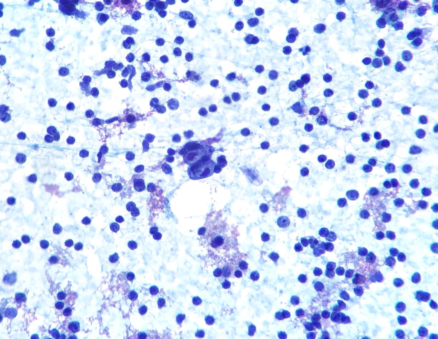
00:01 Our topic now brings us to Hodgkin’s lymphoma. 00:05 Whenever you deal with Hodgkin lymphoma, you should be thinking and contrasting non-Hodgkin’s lymphoma (NHL). 00:13 There are some general features with Hodgkin that do not exist with non-Hodgkin that you need to be quite familiar with. 00:19 And that entire category of NHL or non-Hodgkin’s lymphoma was quite important for us. 00:27 With Hodgkin, it presents in a single lymph node or several adjacent lymph node. 00:33 The type of spread that you find with Hodgkin is called contiguous. 00:36 Non-Hodgkin’s lymphoma, it will be non-contiguous, more common. 00:42 In Hodgkin, it's confined to lymph node with little extranodal component. 00:47 Non-Hodgkin lymphoma, you’d find there to be extranodal involvement to be quite common. 00:54 In Hodgkin, spreads in a contiguous fashion. 00:58 What does that mean? From one node to adjacent lymph node. 01:01 Maybe from the mediastinum to the cervical lymph node. 01:04 However with non-Hodgkin, it will be non-contiguous. 01:09 With Hodgkin, we have absolutely the most important point, histologically you must find a particular B-cell lymphoma or B-cell, excuse me. 01:18 In that particular B-cell, I will show you patterns, morphologically, of Reed-Sternberg cell. 01:25 In pathology, we call this owl eyes. 01:28 And when the time is right, I’ll walk you through different types of Reed-Sternberg. 01:33 If you do not find Reed-Sternberg cell, either in description or upon histology, you cannot diagnose your patient with Hodgkin. 01:45 Etiologies: Reed-Sternberg cells show evidence of EBV infection oftentimes. 01:51 Well, I will walk you through different types of Hodgkin, and I will also give you what the most common type would be. 01:57 One would be called nodular sclerosing and the other type would be mixed cellular. 02:02 Our focus will be on those 2. 02:04 Your focus really should be on nodular sclerosing as you shall see. 02:08 But there is evidence of EBV, very much being involved with Hodgkin. 02:13 The surrounding inflammatory stroma results from expression of cytokines. 02:17 So here from the Reed-Sternberg cell which is technically B-cell, you would expect B-cells to release cytokines and perhaps TNF. 02:25 No exception here. 02:26 You have a cancer cell, a Reed-Sternberg cell, which is behaving like a B-cell but it is pathological. 02:38 An important table for you to understand your different types of Hodgkin lymphoma. 02:44 Subtype nodular sclerosis would be the most common. 02:47 Hence, you pay attention to this. 02:49 Now, before we move on, where is my cancer originating from? From the lymph node. 02:56 Anytime you see the word lymphoma, non-Hodgkin lymphoma or Hodgkin lymphoma, the origin of the cancer will be from the lymph node. 03:05 Is there a possibility that it might enter the circulation? Oh, absolutely. 03:09 Absolutely, so therefore you might have a leukemic type of picture. 03:13 Therefore, at this point, since we’re getting close to completing discussion of Hodgkin lymphoma that all lymphomas will begin/originate from the lymph node as being cancerous and may look leukemic. 03:29 And our discussion of leukemia will be one in which the origin of those cancer and leukemia would be the bone marrow and could enter lymph node. 03:37 Keep that in mind. 03:38 The behavior could be rather similar in presentation. 03:42 However, how it got there, that’s important. 03:46 Now when you say sclerosing, what is undergoing sclerotic process? The lymph node. 03:52 So therefore the lymph node in your head, at this point, really it will be in your best interest to know what a normal lymph node looks like. 04:00 And what happens here is that the lymph node will then undergo sclerosing. 04:03 In other words, narrowing or scarring or in other words, there is more or less fibrotic type of change. 04:11 You lose the normal architecture of the lymph node. 04:14 I will show you different types of Reed-Sternberg cell. 04:17 At this point, I would recommend that you memorize lacunar and classic type of Reed-Sternberg. 04:24 Let me give you a brief description. 04:26 Lacunar, you would expect there to be quite a bit of space within the cell, okay? Wide vacuoles or enlarged vacuoles, lacunar type. 04:36 However, you still would have to find these owl eyes and I will show you those coming up. 04:41 The background here, bands of fibrosis, fibrosis, fibrosis. 04:44 That’s where your focus should be. 04:46 And by fibrosis, I mean the lymph node is becoming fibrosed. 04:50 When it does, what then happens to the actual structure? As you would expect with fibrosis, contracture. 04:57 And so therefore, this will then refer to as being sclerosing. 05:00 Those are the 2 big points, architecture in the Reed-Sternberg and the types. 05:06 Now, the third big point will be the clinical feature. 05:08 Most of your Hodgkin lymphoma, in fact, will be affecting males, males, males. 05:14 Nodular sclerosing is an exception where you will find here that not that there are more females that have greater preponderance over males. 05:24 it’s just the fact that men and women are equally at risk of developing Hodgkin lymphoma. 05:31 Young adults, and I will talk to you about staging in great detail I have to. 05:35 Pay attention to stage I and II. 05:37 That will make more sense to you as I go through a particular classification that will be responsible for every stage as we go through them. 05:46 Mediastinal lymph nodes often times are involved. 05:49 And if the lymph nodes are involved, understand that this is a cancer, so therefore, these lymph nodes will be nontender. 05:56 I'll spend some time with nodular sclerosing more than I would with any other type here. 06:01 Because it is the most common. 06:03 Once you get past this, then you take out a few notes from the remaining types of Hodgkin lymphoma. 06:11 We have mixed cellularity. 06:13 M – mixed cellularity, M – mononuclear. 06:17 Use that to your advantage. 06:18 I will show you a picture of a Reed-Sternberg cell that will be mononuclear type. 06:23 The background here will be mixed, mixed cellularity. 06:26 And here, once again, we’ll get back to the normal preponderance in men or men being more affected than women. 06:35 All ages here, not so much young and look for mixed particularly in the elderly. 06:41 And that’s important. 06:43 And also association with EBV. 06:46 And then I will talk to you about stage III and stage IV. 06:49 Keep in mind, staging always means invasion in pathology. 06:53 And stage IV will be one in which -- Well, now, the cancer, it tends to do what? Metastasize. 07:00 Your third type, now what I wish to bring to your attention is something here called rich. 07:05 All right, this is a lymphocyte-rich. 07:08 Many times, students get this confused with predominant and I could see as to why. 07:11 Because ultimately, in English, by definition, they mean a lot. 07:16 However, on your boards and on the wards, rich, lymphocyte-rich type of Hodgkin lymphoma will be completely different than what’s coming up next, which we’ll talk about as being lymphocyte predominant. 07:30 Keep that in mind and I'll reinforce that when we get to the topic. 07:35 Now the type of Reed-Sternberg cell here will be, once again, mononuclear. 07:38 Then here, it will be T lymphocytes as being background, lymphocyte-rich. 07:43 Uncommon. 07:44 Men once again more so than women that are affected. 07:48 We have lymphocyte depleted, you have a classic type. 07:51 Diffuse perhaps fibrosis, rare, older males and perhaps immunocompromised HIV, lymphocyte depleted. 07:59 So we went from lymphocyte-rich to lymphocyte depleted. 08:02 And then, here is the one that I was referring to, where by definition it sounds like it’s a lot, but be careful though. 08:10 These are 2 different types of Hodgkin. 08:13 We have the rich, as you see here. 08:15 And we have the predominant. 08:17 So this is nodular lymphocyte predominant type. 08:21 Here, the type of Reed-Sternberg cell, and I will show you a picture of this as well, is called popcorn or used to be called popcorn. 08:27 But you have to know this as being L&H type of cell, which stands for lymphocytic and histiocytic type of Reed-Sternberg cell. 08:34 The background here will be more so, a B-cell type. 08:37 Uncommon yet, once again, it would be men that are affected more so than women and it will be the young. 08:43 And it will be cervical axillary versus mediastinal that we find with sclerosing. 08:48 Here are the 5 different types of Hodgkin lymphoma. 08:51 By far, the most common will be sclerosing type. 08:54 Take a few pointers out of the rest so that you’ll be able to distinguish one from the other. 08:59 Understand that you know as to which one of these will be most likely immunocompromised with HIV. 09:04 Well that’s easy, because whenever there is HIV, you’ll know that there is going to be immune status compromise, so lymphocyte depleted. 09:11 And popcorn or L&H will be something with nodular lymphocyte predominant.
About the Lecture
The lecture Lymphadenopathy: Hodgkin Lymphoma (Hodgkin's Disease) – White Blood Cell Pathology by Carlo Raj, MD is from the course Lymphadenopathy – White Blood Cell Pathology (WBC).
Included Quiz Questions
What is the usual presentation of Hodgkin’s lymphoma?
- Multiple lymph nodes in a single region
- Single lymph node along with extranodal component
- Multiple lymph nodes along with excessive extranodal components
- Multiple lymph nodes along with fewer extranodal components
Which of the following is a histological feature of nodular sclerosis lymphoma?
- Partially nodular growth pattern, with fibrous bands separating the nodules
- Abundant RS cells
- Very low lymphocytic count
- Early hematogenous spread
- Never associated with areas of necrosis
Which subtype of Hodgkin’s lymphoma shows dendritic cells?
- Nodular lymphocyte predominant
- Lymphocyte-depleted
- Nodular sclerosis
- Lymphocyte-rich
- Mixed cellularity
Which subtype of Hodgkin’s lymphoma shows popcorn cells?
- Nodular lymphocyte predominant
- Nodular sclerosis
- Mixed cellularity
- Lymphocyte-rich
- Lymphocyte-depleted
Hodgkin's lymphoma with mixed cellularity is commonly detected in which stage?
- Stage III/IV
- Stage II
- Stage I
- Stage 0
- Stage V
Author of lecture Lymphadenopathy: Hodgkin Lymphoma (Hodgkin's Disease) – White Blood Cell Pathology

Carlo Raj, MD
Customer reviews
5,0 of 5 stars
| 5 Stars |
|
2 |
| 4 Stars |
|
0 |
| 3 Stars |
|
0 |
| 2 Stars |
|
0 |
| 1 Star |
|
0 |
Good delineation with the vocabulary terms, lymphocyte rich versus predominant.
well explained this video was good Dr Paul Moss never mentioned types of HL
