Playlist
Show Playlist
Hide Playlist
Development Defects of Conotruncal Ridges

-
Slides 06-31 Formation of the Outflow Tracts.pdf
-
Download Lecture Overview
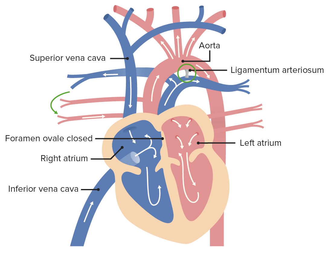
00:01 Now, problems that can occur you can have a persistent truncus arteriosus. 00:06 This occurs when the conotruncal ridges fail to develop and we have a single common outflow tract, so even though there's an aorta and a pulmonary trunk the space leading up to them is one common chamber. 00:19 At this point, you have to have a ventricular septal defect; otherwise, you would not be able to get blood out of one or other of the ventricles. 00:27 So we have severe cyanosis because we have mixing of blood from the left side and the right side. 00:34 Another problem is called transposition of the great vessels. 00:38 This occurs when the conotruncal ridges do form, but instead of spiraling they just go straight up and down and it connect the wrong ventricle to the wrong vessel. 00:48 So the aorta in this case would receive blood from the right ventricle and the pulmonary trunk from the left ventricle. 00:55 If you'd think through how this process is going to work, you'll realize this is absolutely incompatible with life outside the womb because in this situation blood from the body comes to the right atrium, goes to the right ventricle, goes to the aorta and just keeps on recycling without ever getting oxygenated. 01:14 Likewise, blood from the lungs comes into the left atrium, left ventricle and back out to the pulmonary trunk to the lungs again. 01:22 So if you have complete separation of the right and left circuits of the heart, you are unable to survive. 01:28 What happens in these cases when children are born with transposition is there's going to be a shunt, either an atrial septal defect, a ventricular septal defect, a patent foramen ovale or a persistent ductus arteriosus. 01:43 Now, a ductus arteriosus is a structure we're going to devote a lot more time to when we talk about the formation of the vasculature. 01:52 But essentially, it's a connection between the aorta and pulmonary trunk that allows blood to bypass the lungs during embryonic development, to go through the pulmonary trunk and then enter the aorta. 02:03 Once we're born that duct will close up, but if you have transposition you absolutely need to have a shunt like the ductus arteriosus to allow mixing of blood so that you at least have some oxygenated blood making it to your system. 02:18 One very well-described malformation of the conotruncal ridges is called Tetralogy of Fallot. 02:26 Tetralogy because it has four hallmarks, Fallot because Fallot is the person who described it. 02:32 This happens when the conotruncal ridges instead of separating into two equal compartments, it have a very small pulmonary opening and a very large aortic opening. 02:42 So this pulmonary stenosis is the first sign of Tetralogy of Fallot. 02:47 Second sign's that we have a ventricular septal defect and we have to have one because the aorta is so huge and it overrides the ventricular septum. 02:59 The aorta will no longer fit just in the left ventricle so it has to share part of the right ventricle and straddle that ventricular septum. 03:09 The last of four hallmarks is that the right side of the ventricle has to get very big, so the right ventricle enlarges because it's trying its best to push blood through that narrow stenotic pulmonary trunk and it keeps enlarging and getting bigger to try to push harder against that narrowing. 03:29 So when x-rays are taken either anterior/posterior or posterior/anterior, it has a typical boot shape to the heart because of the enlargement of the right ventricle. 03:40 So that's a nice little word association, boot-shaped heart is very commonly used to describe Tetralogy of Fallot. 03:47 So children with this malformation are also known to have what we call tet spells. 03:52 When they're very active they'll become quickly and acutely cyanotic and have to stop what they're doing and squat down. 04:00 That squatting is going to decrease venous return from the lower limbs, so the preload is decreased. This decreases the right-to-left shunt. 04:11 So squeezing off the blood to the lower limbs by squatting down is going to help them recover and become less cyanotic. 04:18 Right ventricular outflow tract obstructions occur anytime there's difficulty getting blood from the right ventricle into the pulmonary trunk. 04:26 We've already seen one form of this. 04:28 It's relatively severe, that's Tetralogy of Fallot. 04:31 When you have pulmonary stenosis, the right side of the heart has to work very hard to push blood to the lungs. 04:39 Now a milder form of this is simply having a less narrow pulmonary trunk. 04:43 But if it's narrow at all, if it's slightly stenotic, you're going to wind up with problems as the right side of the heart tries to compensate for that narrowness by pushing stronger and trying to get more blood into the lungs. 04:57 Over time, that hypertrophy of the ridge of the heart can lead to heart failure unless it's treated. 05:02 The most severe form of right ventricular outflow tract obstruction is hypoplastic right heart syndrome. 05:08 Essentially, the right ventricle doesn't form and the left ventricle is receiving blood from both the right and left atria and just trying to pump the blood to both the aorta and the pulmonary trunk. 05:20 This incompatible with life and is very difficult to repair because there's not much to work with in terms of building a ventricular septum or creating a new right heart or right ventricular side of the heart. 05:35 So you're gonna need a heart transplant if you're gonna get past this particular problem. 05:39 Now on the opposite end, we've got left ventricular outflow tract obstruction and this is gonna be due to aortic stenosis. 05:47 So the left ventricle is gonna have to work very hard to pump blood into the aorta. 05:52 So a mild form of this is aortic stenosis and will lead to heart failure if it's not corrected because the left side of the heart has to pump very hard. 05:59 The most severe form of this is not really surprising, it's hypoplastic left heart syndrome and this is where the left ventricle doesn't form and the right ventricle is doing its best to push blood from both atria into both tracts, the aorta and the pulmonary trunk. 06:17 As with the right side of the heart and hypoplastic right heart syndrome, this is incompatible with further life and is gonna necessitate a heart transplant.
About the Lecture
The lecture Development Defects of Conotruncal Ridges by Peter Ward, PhD is from the course Development of Thoracic Region and Vasculature.
Included Quiz Questions
Which congenital heart defect arises from the failure of the conotruncal ridges to develop properly?
- All of the above
- Persistent truncus arteriosus
- Transposition of the great vessels
- Tetralogy of Fallot
Which congenital heart defect arises due to failure to spiral the formed conotruncal ridges?
- Transposition of the great vessels
- Persistent truncus arteriosus
- Ventricular septal defect
- Atrial septal defect
- Tetralogy of Fallot
Which of the following is not a hallmark finding in tetralogy of Fallot?
- Atrial septal defect
- Pulmonary stenosis
- Ventricular septal defect
- Overriding aorta
- Right ventricular hypertrophy
What is the classical radiologic finding in tetralogy of Fallot?
- Boot-shaped heart
- Egg-on-a-string sign
- Snowman sign
- Scimitar sign
- Gooseneck sign
What is the most serious type of right ventricular outflow tract obstruction?
- Hypoplastic right heart syndrome
- Tetralogy of fallot
- Pulmonary atresia
- Pulmonary valve stenosis
- Truncus arteriosus
What is the most serious type of left ventricular outflow tract obstruction?
- Hypoplastic left heart syndrome
- Aortic valve stenosis
- Supravalvular aortic stenosis
- Coarctation of the aorta
- Bicuspid aortic valve
Author of lecture Development Defects of Conotruncal Ridges

Peter Ward, PhD
Customer reviews
5,0 of 5 stars
| 5 Stars |
|
5 |
| 4 Stars |
|
0 |
| 3 Stars |
|
0 |
| 2 Stars |
|
0 |
| 1 Star |
|
0 |
