Playlist
Show Playlist
Hide Playlist
Cervical Cancer: Risk Factors

-
Slides Cervix Female Reproductive Pathology.pdf
-
Download Lecture Overview
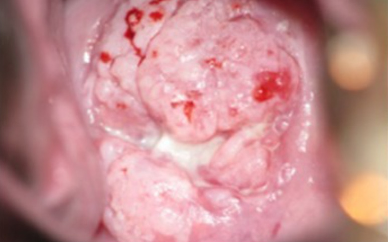
00:01 Let’s talk about cervical cancer and, in verbiage highlight the things we’ve noted. 00:08 E6 and E7. E6 knocks out p53. 00:12 E7 will knock out Rb. 00:15 These are viral oncogenes found in abnormal cervical tissue that has been infected by human papillomavirus known as HPV. 00:22 The E6 and E7 gene proteins transform cells due to their interactions iwth two intracellular proteins called p53 and Rb, which stands for retinoblastoma but in this instance is related to cervical cancer. 00:37 If you have a -- Now, understand the language. 00:41 If you know your basic neoplasia, then you understand that if you have a hypophosphorylated Rb, that’s a key importance, if you have a hypophosphorylated Rb. 00:54 This means that an Rb has not been phosphorylated. 00:57 This means that the Rb is now complexed with E2F. 01:00 And therefore, where is my cell? The cell is stuck between G1/S phase. 01:06 However, if you have E7, the E7 is going to then remove the break. 01:13 So it promotes proteolysis of hypophosphorylated Rb. 01:16 Guess what E7 did. 01:19 It got rid of Rb. 01:20 It removed the break. 01:21 If you remove the break, then E2F allows for a cell to go from G1 to S phase. 01:27 Understand the normal first that I just explained. 01:29 And then what happens here with high grade HPV that possess E6, E7? An issue results in cervical intraepithelial neoplasia. 01:40 In case you missed what CIN stands for, there it is. 01:43 C – cervical, I – intraepithelial, N – neoplasia. 01:48 Is this cancer? No. 01:53 Is this dysplasia? Yes. 01:57 So CIN, precancerous lesion detected by Pap smear. 02:03 Next, the Pap smear has been done, and screening. 02:08 How important has this been for the United States or in the world in general? Really important, right? So therefore, because of the introduction of Pap smear as a screening method, and also vaccination, cervical cancer in developed countries starts dropping in terms of death by gynecologic cancers. 02:33 Worldwide though, cervical cancer is still very, very common. 02:37 The picture, histologically that you’re seeing here, would be an abnormal, abnormal Pap smear, in which you would then expect to find your HPV infected cells with atypical nuclei. 02:50 Once again here, we’ll walk through the major histologic changes. 02:54 Normal on your left, what kind of cells are these? Exocervix, and we have squamous. 03:03 In the middle, I want you to compare the left to the right. 03:07 I want you to focus upon the dysplasia in CIN 3. 03:11 What’s dysplasia mean? Disordered proliferation. 03:16 Lots of proliferation taking place. 03:19 Thick, thick, thick, thick, thick. 03:21 I want you to take a look at the bottom of this. 03:24 That's the basement membrane, that’s the stage. 03:26 Do you see it being ruptured? No. 03:29 This is CIN 3. 03:31 You’re one step closer to? Invasive cervical cancer. 03:37 I want you to compare the basement membrane in CIN 3, which is the bottom of the thickened dysplasia. 03:44 And note, on the right that the membrane has now ruptured and the cancer cells from above are then penetrating and invading into the cervical mucosa. 03:57 Welcome to, unfortunately, a patient who’s developed invasive cervical cancer. 04:05 What then happens after invasion? Here’s your cervical cell cancer. 04:12 You’ll notice here, let me set up the picture, that you've done a pelvic exam, you’re looking at the cervical os. 04:19 But this time, you see mass-like structure sticking out of the cervical os. 04:25 This is not the vagina. The vagina has been removed. 04:28 We’re looking at the cervical os. 04:30 Along with this, what’s behind it is the uterus. 04:34 Okay. 04:35 This mass-like structure that you’re seeing by the cervix, you look at this, no doubt, it’s cancer. 04:40 Upon Pap smear, you'd find your keratin pearls and so on and so forth, then you know it, squamous cell cancer. 04:47 Now, this is a beautiful picture to illustrate. 04:50 What if you start having lateral invasion of your cervical cancer? You can now close your eyes. 04:57 Think about the cervix and the surrounding structures that you have If you start laterally invading, and your patient starts showing signs of changes in BUN and creatinine. 05:10 And you find your BUN creatinine ratio to be elevated. 05:15 Elevated. 05:16 "What does that has to do with your cervix, Dr. Raj?" BUN creatinine is a measurement of your kidney. Yes But if you have lateral invasion in the cervix, what structure are you then obstructing? There you go. 05:29 You’re obstructing the kidney. 05:32 Your patient, often cause of death is post renal failure because of lateral invasion of your cancer in cervical cancer.
About the Lecture
The lecture Cervical Cancer: Risk Factors by Carlo Raj, MD is from the course Disorders of Vulva, Vagina and Cervix.
Included Quiz Questions
Which HPV proteins cause mutations in oncogenes leading to malignant changes in the cervical epithelium?
- E6 and E7
- E4 and E6
- E7 and E9
- E16 and E18
- E5 and E6
The integrity of what structure differentiates between high-grade intraepithelial lesion (carcinoma in situ) and invasive epithelial carcinoma?
- Basement membrane of the epithelium
- Endothelium of the closest blood vessel
- Fascia between the epithelium and the muscle layer
- Squamocolumnar junction
- Peritoneum
What is the MOST effective measure for early detection and decreased mortality from cervical cancer?
- Regular Pap smears
- Screening for serum anti-HPV antibodies
- Mandatory education about contraceptive use
- Annual screening for sexually transmitted infections
- Routine childhood vaccinations
Author of lecture Cervical Cancer: Risk Factors

Carlo Raj, MD
Customer reviews
5,0 of 5 stars
| 5 Stars |
|
1 |
| 4 Stars |
|
0 |
| 3 Stars |
|
0 |
| 2 Stars |
|
0 |
| 1 Star |
|
0 |
Awesome presentation. The lecture was precise and straight to the point, with quality pieces of information.
