Table of Contents
Image: “Medical Students.” by Ximehernandez1409. License: CC BY-SA 3.0
Definition: What is Histology?
Histology is a component of microscopic anatomy
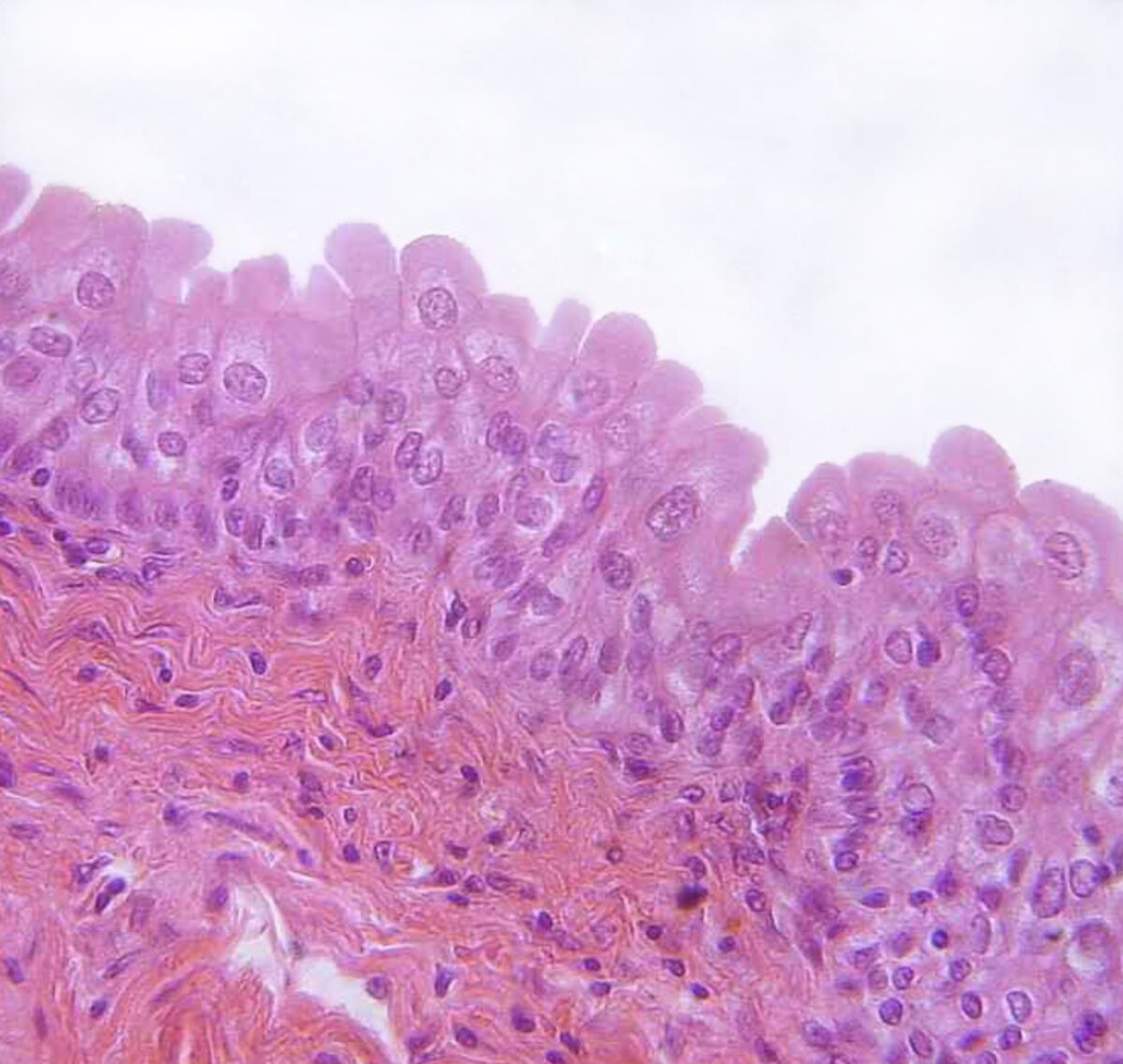
Histology (Greek: histos = tissue and logos = teaching) is the study of tissues. It belongs to the field of microscopic anatomy. It deals with the arrangement and structure of cells and their organization as a cellular unit in various tissue types.
Histology curriculum and a microscopy course are offered in the 1st and 2nd semesters of preclinical studies, which are especially relevant for the subsequent clinical phase, as they provide the requisite diagnostic skills based on tissue studies.

Image: Slice preparation for light microscopy: 1 glass slides (76 x 26 mm), 2 cover glass, in between: 3 colored organ section. By Rollroboter, License: CC BY-SA 3.0
General Histology
The 1st step entails identifying the specimen via direct observation, i.e., without a microscope, to determine the number of edges and any lumen visible.
The specimen is viewed under the lowest magnification, in order to obtain a general overview and develop a hypothesis eventually. The magnification can be increased steadily to capture the details, and thus confirm or disprove the hypothesis.
For orientation purposes, it is advisable to always return to the lowest magnification and repeatedly zoom in on the parts of interest. However, for a successful histology lesson, in addition to the identification of the tissue specimen, thorough background knowledge is essential.
Tissues
1. Epithelial tissue
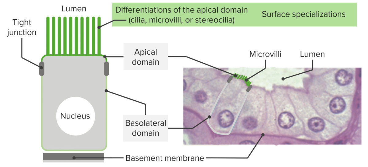
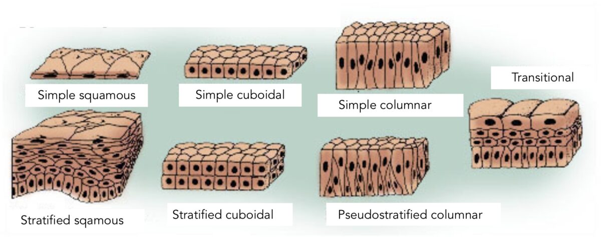
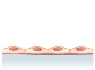
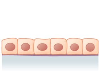
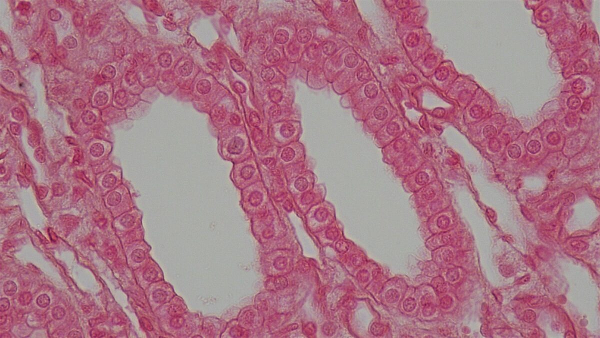
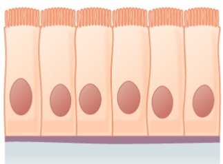
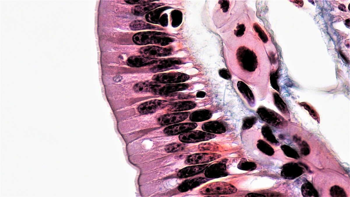
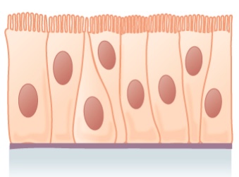
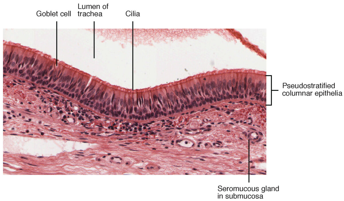
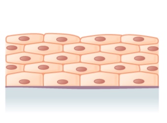
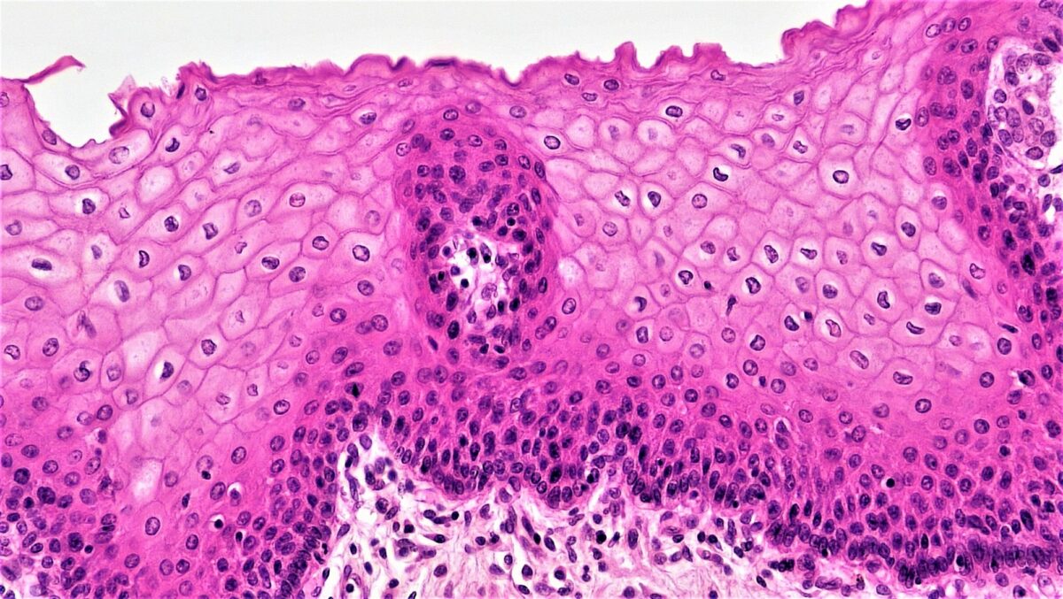
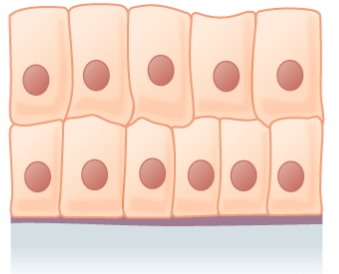
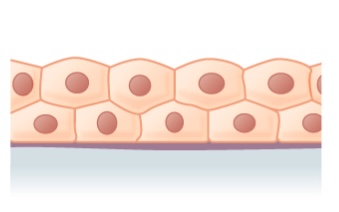
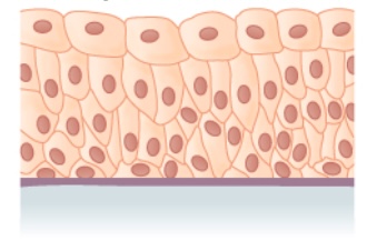
Epithelium represents a group of cells that line various organs. The 3 principal shapes include:
- Squamous
- Cuboidal
- Columnar
The presence of epithelium provides an important clue for tissue diagnosis. The type of epithelium further facilitates the identification of the tissue section.
In addition to the systematic structure, the distribution and function of all epithelial types are essential for the understanding of pathology.
2. Bone tissue
3. Muscle tissue
Muscles define the body; therefore, it is vital to understand muscle function. Movement is the result of alternating contraction and relaxation of muscles, which account for approx. 30–40% of the total body weight. Muscles are primarily engaged in the conversion of chemical energy into mechanical energy to perform work.
Histologically, the muscles of the body can be classified into 3 different types:
Smooth muscles as visceral muscles (muscles of internal hollow organs) | ![]() |
Cardiac muscle or myocardium | ![]() |
Striated or skeletal muscle as a component of active musculoskeletal system | ![]() |
Image: Partial View of ‘the musculature of our body can be differentiated into 3 types’. By Phil Schatz, License: CC BY 4.0
A concise overview of the different types of muscle, their structure, their movement from their origin and insertion points, and the auxiliaries used are essential for any physician.
Cells
The cell is the functional unit of a living organism. Organisms such as bacteria and algae are unicellular while complex organisms such as human beings are multicellular. In addition, complex organisms contain an extracellular matrix and exhibit complex intracellular structures.
The cell is the basic building block of all life, measuring only a few microns in size. Nevertheless, it accomplishes incredible feats as a biological unit. The numerous organelles, the cytoskeleton, and cell-cell junctions are popular topics in biology, biochemistry, and histology examinations.
Special Histology
Which organs contain which tissue types and in what composition and how do these cell groups function as a whole?
Blood
Blood (Latin: sanguis, Greek: haima) is a suspension of various cells in a protein-containing saline solution. It accounts for 6–8% of body weight and has a pH value of 7.4. Blood performs numerous vital functions and is the key player in the entire organism.
Blood components (%) | Blood subcomponents (%) | Type (%) | Production site | Main functions |
Plasma (43–63%) | Water (92%) | Liquid | Absorbed through the intestinal tract or synthesized during metabolism | Transport medium |
Plasma proteins (7%) | Albumin (54–60%) | Liver | Maintenance of osmotic concentration, and transport of lipid molecules | |
Globulin (35–38%) | Alpha globulin—Liver | Transport, maintenance of osmotic concentration | ||
Beta globulin—Liver | Transport, maintenance of osmotic concentration | |||
Gamma globulin (Immunoglobulin)—Plasma cells | Immune response | |||
Fibrinogen (4–7%) | Liver | Coagulation during hemostasis | ||
Regulatory proteins (< 1%) | Hormones and enzymes | Various sites | Regulation of various body functions | |
Other solutes (1%) | Nutrients, gases, and waste products | Intestinal absorption, gas exchange in the respiratory tract, or cellular synthesis | Several different functions | |
Main components (37–54%) | Erythrocytes (99%) | Erythrocytes | Red bone marrow | Gas transport, primarily oxygen and carbon dioxide |
Leucocytes (< 1%), thrombocytes (< 1%) | Granulocytes: neutrophils, eosinophils, and basophils | Red bone marrow | Innate immunity | |
Agranular leukocytes: lymphocytes, monocytes | Lymphocytes: bone marrow and lymphatic tissue | Lymphocytes: adaptive immunity |
||
Monocytes: red bone marrow | Monocytes: innate immunity | |||
Thrombocytes (< 1 %) | Megakaryocytes: red bone marrow | Coagulation |
Blood vessels: The wall structure of arteries and veins
Blood vessels move in and out of a histological section. They should be recognized as such during the differentiation of arteries from veins. The main difference lies in the wall structure.
Circulatory system
The human circulatory system is the convective transport of respiratory gases, nutrients, hormones, and heat via blood, which is pumped from the heart through the blood vessels and returned to the heart.
The blood circulation changes rapidly but is indispensable for the maintenance of body functions. High blood pressure (arterial hypertension) is one of the most common diseases in society. It is therefore important for physicians to know and understand the circulatory system.
Respiratory system
Along with the intestines and the skin, the lung is another organ in direct contact with the outside world. It facilitates gas exchange by diffusion. Pulmonary gas exchange every second requires a transport system for waste gases and fresh air.
A space in which the walls are thin enough for gas diffusion is as important as a transport system for the gases in a chemically bound form. Histologists must closely analyze the management of these tasks by the individual structures.
The integumentary system
The skin, subcutaneous fat, hair, and nails protect us from hazardous environmental exposures. Towards this end, they must be sealed so that our body fluids are not depleted. The skin is the largest organ and weighs 3–10 kg. In an individual who is 170 cm tall, the surface area of the skin equals 1.8 m². The skin is also the site of vitamin D synthesis.
Kidney and excretory organs
The kidney is a vital excretory organ and controls the water and electrolyte balance in our body. Their precise mechanism of action can only be understood at the microscopic level. Each renal tissue type is designed for a specific purpose, and the sophisticated functions of the kidney can be traced to its histology.
Endocrine organs and hormonal system
In the absence of a hormonal system, our bodies would not function as a whole. Endocrine organs ensure coordinated communication between specialized individual systems for an appropriate and adequate response to environmental factors.
The control of body functions via the endocrine system is the subject of anatomy, physiology, and biochemistry. Even in clinical practice, a comprehensive understanding of the hormones and their functions is essential. Pituitary, thyroid, parathyroid, adrenal, and pineal glands represent endocrine organs, in the strictest sense of the term. Based on their characteristic structure, they are not difficult to identify histologically; however, a few elements should be considered.
Endocrine glands and their primary hormones
Endocrine gland | Associated hormones | Chemical class | Effect |
Pituitary gland (anterior) | Growth hormone | Protein | Promotes the growth of body tissues |
Prolactin | Peptide | Promotes milk production | |
Thyroid-stimulating hormone (TSH) | Glycoprotein | Stimulates thyroid hormone secretion | |
ACTH | Peptide | Stimulates the release of hormones from the adrenal cortex | |
Follicle-stimulating hormone (FSH) | Glycoprotein | Stimulates gametogenesis | |
Luteinizing hormone (LH) | Glycoprotein | Stimulates gonadal androgen production | |
Pituitary gland (posterior) | ADH | Peptide | Stimulates water absorption in the kidneys |
Oxytocin | Peptide | Stimulates uterine contractions during childbirth | |
Thyroid gland | Thyroxine (T4); triiodothyronine (T3) | Amine | Stimulates basal metabolic rate |
Calcitonin | Peptide | Lowers blood Ca2+ levels | |
Parathyroid gland | Parathyroid hormone | Peptide | Raises blood Ca2+ levels |
Adrenals (cortex) | Aldosterone | Steroid | Raises blood Na2+ levels |
Cortisol, corticosterone, cortisone | Steroid | Raises blood sugar levels | |
Adrenals (medulla) | Adrenaline, noradrenaline | Amine | Stimulates fight or flight response |
Pineal | Melatonin | Amine | Controls the sleep cycle |
Pancreas | Insulin | Protein | Lowers blood sugar levels |
Glucagon | Protein | Raises blood sugar levels | |
Testes | Testosterone | Steroid | Stimulates the development of male secondary sexual characteristics and sperm production |
Ovaries | Estrogen and progesterone | Steroid | Stimulate the development of female secondary sex characteristics and prepares the body for childbirth |
Table: Endocrine glands and their major hormones. By Phil Schatz, License: CC BY 4.0
The immune system
The lymphoid organs, which include the thymus, spleen, lymph nodes and tonsils, are exciting points of interest, and important topics in medicine. The lymphatic system plays a role in numerous physiological events throughout the body, especially in the complex defense mechanisms. Understanding the lymphatic system is crucial for the diagnosis of several pathologies.
Nervous system
The nervous system ‘only’ weighs 2 kg, which amounts to approx. 3% of the total body weight. It is a small but complex network of neurons and an even greater number of neuroglia. For all physicians, it is important to know the structure and physiology of the nervous system.
Sensory system
The sensory organs allow us to interact with our environment and to perceive things outside of our bodies. Their functions are fascinating and complex and are substantial topics in the study of medicine.
Despite the sheer diversity of the sense organs, they conform to basic principles of structure and function.
Digestive system and gastrointestinal tract

Image: Digestive system with liver. By Gordon Flynn, License: CC BY-SA 2.5
The digestive system is an immense challenge given the vast but subtle differences between the individual components.
The breakdown of nutrients and their assimilation depend on digestion. Nutrients are then transported into individual cells of the body via the bloodstream.
Pancreas
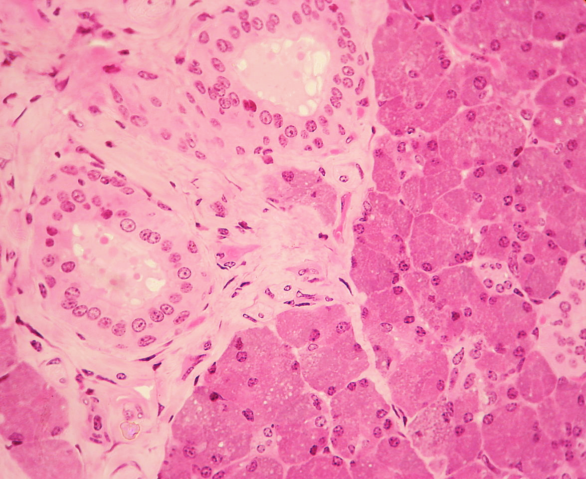
The pancreas is both an exocrine and an endocrine organ. It is essential for digestion and metabolism of carbohydrates, which is why a loss of function leads to severe diseases.
Female Reproductive System—follicular, mucous and breast tissue
The female sex organs undergo periodic and recurring structural changes. In female sex organs, a number of physiological events occur under hormonal influence. Topics such as folliculogenesis in the uterus, the structure of the mammary tissue, and lactation are important from a medical perspective.
Male reproductive system
The understanding of the male reproductive system requires a comprehensive biological insight. The male reproductive organs comprising testis and vas deferens are composed of different sections.
Histological Stains
Staining is an additional microscopic procedure that enhances the contrast of the microbiological image to highlight tissues in focus. The different histological stains and dye stains used are discussed here.
Hematoxylin and Eosin (H&E) stain

Image: Islet of Langerhans, haemalum-eosin stain. By Polarlys, License: CC BY-SA 3.0
Azan stain

Image: Modified stained with azan: collagenous connective tissue blue. epididymis. By Rollroboter, License: CC BY-SA 3.0
Elastica stain

Image: Elastic fibers in mammary glands colored by Weigert’s elastic stain. Elastic fibers are blue. By R.Denzer, License: CC BY-SA 3.0
Van Gieson’s stain

Image: Colored bronchi of ostrich, after Van Gieson. By Lycaon.cl, License: CC BY-SA 4.0
Crossmon stain
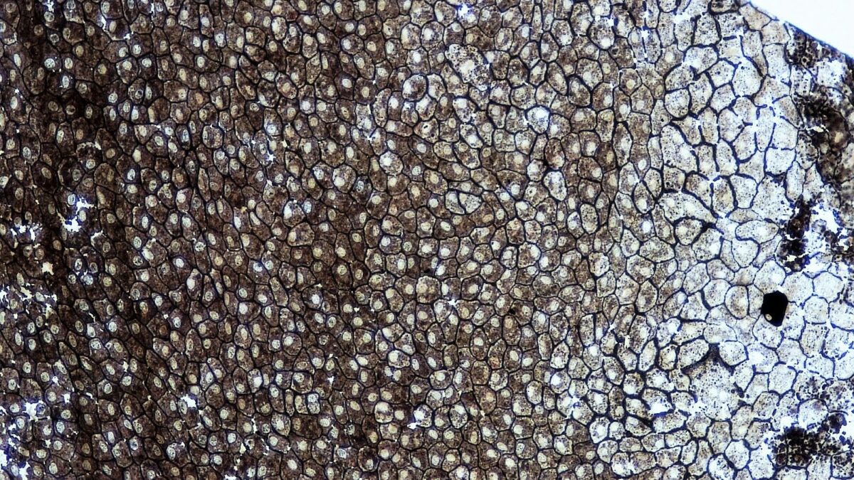
The Crossmon stain consists of iron hematoxylin, orange G and light green SF. It can be used to stain the nucleus black-brown, while the cytoplasm appears reddish. Collagen fibers take on a green hue. Elastic fibers and bones are light-red. Erythrocytes are stained orange with Crossmon.
Special stains
A number of histochemical stains provide a glimpse into dynamic cellular events. For instance, carbohydrate components such as glycoproteins or glycogen (PAS reaction), negatively charged components such as glycosaminoglycans or hyaluronic acid (Alcian blue stain), or lipids (fatty dyes such as Sudan III) represent the site of their natural location within cells and tissues.
Finally, immunohistochemical methods are used to detect specific proteins or peptides based on antigen-antibody reaction.
In-situ hybridization is the method of choice to identify specific DNA or RNA sequences. Radioactively or non-radioactively labeled samples are used to localize the histological specimens appropriately.
Study Table: Commonly used stains
Stain | General application | Nucleus | Cytoplasm | Red blood cells | Collagen fibers | Special stains |
Hematoxylin | Commonly used stain, often paired with eosin | Orange, cyan, blue or green | Blue/brown /black | N/A | N/A | Nucleic acids: blue; endoplasmic reticulum: blue |
Eosin | Commonly used stain, often paired with hematoxylin | N/A | Pink | Orange/red | Pink | Elastic fibers: pink; collagen fibers: pink; reticular fibers: pink |
Toluidine blue | Commonly used stain | Blue | Blue | Blue | Blue | Mast cell granules: purple |
Masson’s trichrome stain | Connective tissue | Black | Red/Pink | Red | Blue/Green | Cartilage: blue/green; muscle fibers: red |
Mallory´s trichrome stain | Connective tissue | Red | Pale red | Orange | Dark blue | Keratin: orange; cartilage: blue; bone matrix: dark blue; muscle fibers: red |
Elastica stain | Elastica fibers | Blue/black | N/A | N/A | N/A | Elastic fibers: blue/black |
Azan stain | Separates cells from extracellular components | Red/purple | Pink | Red | Blue | Muscle fibers: red; cartilage: blue; bone matrix: blue |
Silver stain |
Reticular fibers, nerve fibers, fungi | N/A | N/A | N/A | N/A | Reticular fibers: brown/black; nerve fibers: brown/black; fungi: black |
Wright stain |
Blood cells | Bluish/purple | Bluish/purple | Red/Pink | N/A | Neutrophil granules: purple/pink; eosinophil granules: bright red/orange; basophil granules: dark purple/violet; thrombocyte granules: red/purple |
Orcein stain | Elastic fibers | Dark blue | N/A | Light red | Pink | Elastic fibers: dark brown; mast cell granules: purple; smooth muscle: light-blue |
PAS reaction | Basal membrane, localization of carbohydrates | Blue | N/A | N/A | Pink | Glycogen and other carbohydrates: magenta |
Table: Common laboratory stains. By Wikipedia.
Histology is a subject central to medicine and covers areas of anatomy, biology, physiology, even through to biochemistry. A basic understanding of histology facilitates the learning of other essential subjects to appreciate pre-clinical findings covering not only individualized compartments but the tissue as an integrated whole.
Histology mnemonics:
- Titin sounds like Titan and is the largest protein in the body.
- T-cells come from the thymus.
- B-cells come from the bone.
- Plexus submucosus (Meissner) is responsible for the mucosa centrally.
- Plexus myentericus (Auerbach) innervates muscles externally.
- Osteoclasts chew up bone.
- Osteoblasts build the bone.
- C-cells synthesize calcitonin, which reduces Ca2+-levels in plasma.
- Parathyroid hormone provides the body with Ca2+ (in blood).
Leave a Reply