Achieve Mastery of Medical Concepts

Study for medical school and boards with Lecturio

Gestational Trophoblastic Disease

Gestational trophoblastic diseases are a spectrum of placental disorders resulting from abnormal placental trophoblastic growth. These disorders range from benign Benign Fibroadenoma molar pregnancies (complete and partial moles Moles Primary Skin Lesions) to neoplastic conditions such as invasive moles Moles Primary Skin Lesions and choriocarcinoma. Diagnosis is confirmed by elevated serum beta human chorionic gonadotropin (hCG) and ultrasound findings, which are dependent on the disorder. Treatment is primarily through dilation and curettage Curettage A scraping, usually of the interior of a cavity or tract, for removal of new growth or other abnormal tissue, or to obtain material for tissue diagnosis. It is performed with a curet (curette), a spoon-shaped instrument designed for that purpose. Benign Bone Tumors and/or methotrexate Methotrexate An antineoplastic antimetabolite with immunosuppressant properties. It is an inhibitor of tetrahydrofolate dehydrogenase and prevents the formation of tetrahydrofolate, necessary for synthesis of thymidylate, an essential component of DNA. Antimetabolite Chemotherapy.

Last updated: Oct 24, 2022

Editorial responsibility: Stanley Oiseth, Lindsay Jones, Evelin Maza

Overview

Epidemiology

  • Occurs in about 1 pregnancy Pregnancy The status during which female mammals carry their developing young (embryos or fetuses) in utero before birth, beginning from fertilization to birth. Pregnancy: Diagnosis, Physiology, and Care in 1,200 pregnancies in the United States, with hydatidiform mole Mole Nevi (singular nevus), also known as “moles,” are benign neoplasms of the skin. Nevus is a non-specific medical term because it encompasses both congenital and acquired lesions, hyper- and hypopigmented lesions, and raised or flat lesions. Nevus/Nevi representing 80 percent of the cases. Higher incidence Incidence The number of new cases of a given disease during a given period in a specified population. It also is used for the rate at which new events occur in a defined population. It is differentiated from prevalence, which refers to all cases in the population at a given time. Measures of Disease Frequency in Southeast Asia ASIA Spinal Cord Injuries and Japan 
  • Risk factors:
    • History of gestational trophoblastic disease (GTD)
    • History of prior spontaneous abortion Abortion Expulsion of the product of fertilization before completing the term of gestation and without deliberate interference. Spontaneous Abortion and/or infertility Infertility Infertility is the inability to conceive in the context of regular intercourse. The most common causes of infertility in women are related to ovulatory dysfunction or tubal obstruction, whereas, in men, abnormal sperm is a common cause. Infertility
    • Protein, folic acid, and carotene deficiency

Classification

  • Hydatidiform moles Moles Primary Skin Lesions: considered premalignant because of their potential for malignancy Malignancy Hemothorax
    • Complete mole Mole Nevi (singular nevus), also known as “moles,” are benign neoplasms of the skin. Nevus is a non-specific medical term because it encompasses both congenital and acquired lesions, hyper- and hypopigmented lesions, and raised or flat lesions. Nevus/Nevi
    • Partial mole Mole Nevi (singular nevus), also known as “moles,” are benign neoplasms of the skin. Nevus is a non-specific medical term because it encompasses both congenital and acquired lesions, hyper- and hypopigmented lesions, and raised or flat lesions. Nevus/Nevi
  • Invasive moles Moles Primary Skin Lesions
  • Choriocarcinoma: incidence Incidence The number of new cases of a given disease during a given period in a specified population. It also is used for the rate at which new events occur in a defined population. It is differentiated from prevalence, which refers to all cases in the population at a given time. Measures of Disease Frequency USA 1/40,000

Hydatidiform Mole

Hydatidiform moles Moles Primary Skin Lesions are characterized by cystic Cystic Fibrocystic Change swelling Swelling Inflammation of the chorionic villi Chorionic villi Threadlike vascular projections of the chorion. Chorionic villi may be free or embedded within the decidua forming the site for exchange of substances between fetal and maternal blood (placenta). Placenta, Umbilical Cord, and Amniotic Cavity and proliferation of the chorionic epithelium Epithelium The epithelium is a complex of specialized cellular organizations arranged into sheets and lining cavities and covering the surfaces of the body. The cells exhibit polarity, having an apical and a basal pole. Structures important for the epithelial integrity and function involve the basement membrane, the semipermeable sheet on which the cells rest, and interdigitations, as well as cellular junctions. Surface Epithelium: Histology. There are 2 types: complete mole Mole Nevi (singular nevus), also known as “moles,” are benign neoplasms of the skin. Nevus is a non-specific medical term because it encompasses both congenital and acquired lesions, hyper- and hypopigmented lesions, and raised or flat lesions. Nevus/Nevi and partial mole Mole Nevi (singular nevus), also known as “moles,” are benign neoplasms of the skin. Nevus is a non-specific medical term because it encompasses both congenital and acquired lesions, hyper- and hypopigmented lesions, and raised or flat lesions. Nevus/Nevi.

Complete mole Mole Nevi (singular nevus), also known as “moles,” are benign neoplasms of the skin. Nevus is a non-specific medical term because it encompasses both congenital and acquired lesions, hyper- and hypopigmented lesions, and raised or flat lesions. Nevus/Nevi Partial mole Mole Nevi (singular nevus), also known as “moles,” are benign neoplasms of the skin. Nevus is a non-specific medical term because it encompasses both congenital and acquired lesions, hyper- and hypopigmented lesions, and raised or flat lesions. Nevus/Nevi
Karyotype Karyotype The full set of chromosomes presented as a systematized array of metaphase chromosomes from a photomicrograph of a single cell nucleus arranged in pairs in descending order of size and according to the position of the centromere. Congenital Malformations of the Female Reproductive System 46,XX or 46,XY Triploid (69,XXX, 69, XXY XXY Klinefelter syndrome is a chromosomal aneuploidy characterized by the presence of 1 or more extra X chromosomes in a male karyotype, most commonly leading to karyotype 47,XXY. Klinefelter syndrome is associated with decreased levels of testosterone and is the most common cause of congenital hypogonadism. Klinefelter Syndrome, or 69,XYY)
Formed from Enucleated egg and a single sperm 2 sperm and 1 egg
Fetal parts Absent Present
Human chorionic gonadotropin (HCG) level ↑↑↑
Ultrasound findings
  • Honeycomb uterus Uterus The uterus, cervix, and fallopian tubes are part of the internal female reproductive system. The uterus has a thick wall made of smooth muscle (the myometrium) and an inner mucosal layer (the endometrium). The most inferior portion of the uterus is the cervix, which connects the uterine cavity to the vagina. Uterus, Cervix, and Fallopian Tubes: Anatomy
  • “Clusters of grapes”
  • “Snowstorm” uterus Uterus The uterus, cervix, and fallopian tubes are part of the internal female reproductive system. The uterus has a thick wall made of smooth muscle (the myometrium) and an inner mucosal layer (the endometrium). The most inferior portion of the uterus is the cervix, which connects the uterine cavity to the vagina. Uterus, Cervix, and Fallopian Tubes: Anatomy
Reveals fetal parts
Malignancy Malignancy Hemothorax risk Higher risk for choriocarcinoma Rare

Clinical presentation

  • Vaginal bleeding
  • Uterine enlargement (more than expected for given gestational age Gestational age The age of the conceptus, beginning from the time of fertilization. In clinical obstetrics, the gestational age is often estimated as the time from the last day of the last menstruation which is about 2 weeks before ovulation and fertilization. Pregnancy: Diagnosis, Physiology, and Care)
  • Pelvic pressure or pain Pain An unpleasant sensation induced by noxious stimuli which are detected by nerve endings of nociceptive neurons. Pain: Types and Pathways
  • May be associated with 
    • Early (before 20 weeks) preeclampsia Preeclampsia A complication of pregnancy, characterized by a complex of symptoms including maternal hypertension and proteinuria with or without pathological edema. Symptoms may range between mild and severe. Pre-eclampsia usually occurs after the 20th week of gestation, but may develop before this time in the presence of trophoblastic disease. Hypertensive Pregnancy Disorders
    • Theca-lutein cysts Cysts Any fluid-filled closed cavity or sac that is lined by an epithelium. Cysts can be of normal, abnormal, non-neoplastic, or neoplastic tissues. Fibrocystic Change
    • Hyperemesis gravidarum
    • Hyperthyroidism Hyperthyroidism Hypersecretion of thyroid hormones from the thyroid gland. Elevated levels of thyroid hormones increase basal metabolic rate. Thyrotoxicosis and Hyperthyroidism

Diagnosis

  • HCG levels: ↑ serum beta-hCG 
  • Ultrasound:
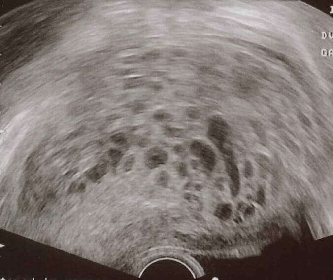
    • Reveals an irregular distribution of hypoechoic (cystic) and hyperechoic (solid) areas within the uterus
    • Characteristically referred to as a “snowstorm pattern
Transvaginal ultrasonography showing a molar pregnancy

Transvaginal ultrasound of a hydatidiform mole: A characteristic “snowstorm pattern” is observed in ultrasound scan.

Image: “Transvaginal ultrasonography showing a molar pregnancy” by Mikael Häggström. License: CC0
Treatment

  • Suction dilation and curettage Curettage A scraping, usually of the interior of a cavity or tract, for removal of new growth or other abnormal tissue, or to obtain material for tissue diagnosis. It is performed with a curet (curette), a spoon-shaped instrument designed for that purpose. Benign Bone Tumors
  • HCG levels are monitored (expected to downtrend after treatment)
  • Hysterectomy (last resort)
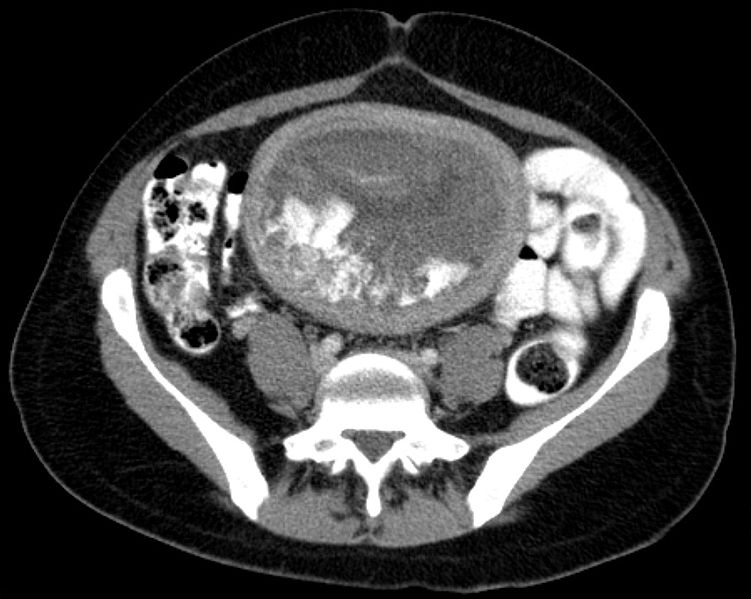
Blasenmole ct axial

Hydatid in axial computed tomography (CT) image

Image: “Blasenmole Computertomographie axial” by Hellerhoff. License: CC BY-SA 3.0

Related videos

Choriocarcinoma

Choriocarcinoma is a highly aggressive malignant neoplasm of trophoblastic cells that can develop during or after pregnancy Pregnancy The status during which female mammals carry their developing young (embryos or fetuses) in utero before birth, beginning from fertilization to birth. Pregnancy: Diagnosis, Physiology, and Care in the mother or baby.

Etiology

Can be preceded by:

  • Hydatidiform mole Mole Nevi (singular nevus), also known as “moles,” are benign neoplasms of the skin. Nevus is a non-specific medical term because it encompasses both congenital and acquired lesions, hyper- and hypopigmented lesions, and raised or flat lesions. Nevus/Nevi (50%)
  • Abortion Abortion Expulsion of the product of fertilization before completing the term of gestation and without deliberate interference. Spontaneous Abortion of an ectopic pregnancy Ectopic pregnancy Ectopic pregnancy refers to the implantation of a fertilized egg (embryo) outside the uterine cavity. The main cause is disruption of the normal anatomy of the fallopian tube. Ectopic Pregnancy (20%)
  • Normal-term pregnancy Pregnancy The status during which female mammals carry their developing young (embryos or fetuses) in utero before birth, beginning from fertilization to birth. Pregnancy: Diagnosis, Physiology, and Care (30%)

Clinical presentation

  • Abnormal uterine bleeding Abnormal Uterine Bleeding Abnormal uterine bleeding is the medical term for abnormalities in the frequency, volume, duration, and regularity of the menstrual cycle. Abnormal uterine bleeding is classified using the acronym PALM-COEIN, with PALM representing the structural causes and COEIN indicating the non-structural causes. Abnormal Uterine Bleeding or amenorrhea Amenorrhea Absence of menstruation. Congenital Malformations of the Female Reproductive System
  • If hematogenous Hematogenous Hepatocellular Carcinoma (HCC) and Liver Metastases spread to the lungs Lungs Lungs are the main organs of the respiratory system. Lungs are paired viscera located in the thoracic cavity and are composed of spongy tissue. The primary function of the lungs is to oxygenate blood and eliminate CO2. Lungs: Anatomy: shortness of breath Shortness of breath Dyspnea is the subjective sensation of breathing discomfort. Dyspnea is a normal manifestation of heavy physical or psychological exertion, but also may be caused by underlying conditions (both pulmonary and extrapulmonary). Dyspnea and/or hemoptysis Hemoptysis Hemoptysis is defined as the expectoration of blood originating in the lower respiratory tract. Hemoptysis is a consequence of another disease process and can be classified as either life threatening or non-life threatening. Hemoptysis can result in significant morbidity and mortality due to both drowning (reduced gas exchange as the lungs fill with blood) and hemorrhagic shock. Hemoptysis

Diagnosis

  • HCG levels: ↑ serum beta-hCG
  • Pelvic ultrasound: hypervascularity
  • Chest X-ray: pulmonary metastases with “cannonball” shape

Treatment

  • Depends on stage and risk classification
  • Low risk: methotrexate Methotrexate An antineoplastic antimetabolite with immunosuppressant properties. It is an inhibitor of tetrahydrofolate dehydrogenase and prevents the formation of tetrahydrofolate, necessary for synthesis of thymidylate, an essential component of DNA. Antimetabolite Chemotherapy monotherapy or combination with actinomycin D Actinomycin D A compound composed of a two cyclic peptides attached to a phenoxazine that is derived from streptomyces parvulus. It binds to DNA and inhibits RNA synthesis (transcription), with chain elongation more sensitive than initiation, termination, or release. As a result of impaired mRNA production, protein synthesis also declines after dactinomycin therapy. Antitumor Antibiotics
  • High risk: multi-agent regimen
    • Etoposide Etoposide A semisynthetic derivative of podophyllotoxin that exhibits antitumor activity. Etoposide inhibits DNA synthesis by forming a complex with topoisomerase II and DNA. This complex induces breaks in double stranded DNA and prevents repair by topoisomerase II binding. Accumulated breaks in DNA prevent entry into the mitotic phase of cell division, and lead to cell death. Etoposide acts primarily in the g2 and s phases of the cell cycle. Microtubule and Topoisomerase Inhibitors
    • Methotrexate Methotrexate An antineoplastic antimetabolite with immunosuppressant properties. It is an inhibitor of tetrahydrofolate dehydrogenase and prevents the formation of tetrahydrofolate, necessary for synthesis of thymidylate, an essential component of DNA. Antimetabolite Chemotherapy ( MTX MTX An antineoplastic antimetabolite with immunosuppressant properties. It is an inhibitor of tetrahydrofolate dehydrogenase and prevents the formation of tetrahydrofolate, necessary for synthesis of thymidylate, an essential component of DNA. Antimetabolite Chemotherapy)
    • Actinomycin D Actinomycin D A compound composed of a two cyclic peptides attached to a phenoxazine that is derived from streptomyces parvulus. It binds to DNA and inhibits RNA synthesis (transcription), with chain elongation more sensitive than initiation, termination, or release. As a result of impaired mRNA production, protein synthesis also declines after dactinomycin therapy. Antitumor Antibiotics
    • Leucovorin calcium Calcium A basic element found in nearly all tissues. It is a member of the alkaline earth family of metals with the atomic symbol ca, atomic number 20, and atomic weight 40. Calcium is the most abundant mineral in the body and combines with phosphorus to form calcium phosphate in the bones and teeth. It is essential for the normal functioning of nerves and muscles and plays a role in blood coagulation (as factor IV) and in many enzymatic processes. Electrolytes
    • Cyclophosphamide Cyclophosphamide Precursor of an alkylating nitrogen mustard antineoplastic and immunosuppressive agent that must be activated in the liver to form the active aldophosphamide. It has been used in the treatment of lymphoma and leukemia. Its side effect, alopecia, has been used for defleecing sheep. Cyclophosphamide may also cause sterility, birth defects, mutations, and cancer. Immunosuppressants
    • Vincristine Vincristine An antitumor alkaloid isolated from vinca rosea. Microtubule and Topoisomerase Inhibitors
Interface between choriocarcinoma with central necrosis and normal placenta

Choriocarcinoma with central necrosis

Image: “Interface between choriocarcinoma with central necrosis and normal placenta” by Department of Obstetrics and Gynecology, St. Louis University, Missouri, USA. License: CC BY 2.0

Clinical Relevance

Ectopic pregnancy Ectopic pregnancy Ectopic pregnancy refers to the implantation of a fertilized egg (embryo) outside the uterine cavity. The main cause is disruption of the normal anatomy of the fallopian tube. Ectopic Pregnancy: Eccyesis Eccyesis Ectopic pregnancy refers to the implantation of a fertilized egg (embryo) outside the uterine cavity. The main cause is disruption of the normal anatomy of the fallopian tube. Ectopic Pregnancy or ectopic pregnancy Ectopic pregnancy Ectopic pregnancy refers to the implantation of a fertilized egg (embryo) outside the uterine cavity. The main cause is disruption of the normal anatomy of the fallopian tube. Ectopic Pregnancy refers to the implantation Implantation Endometrial implantation of embryo, mammalian at the blastocyst stage. Fertilization and First Week of the blastocyst Blastocyst A post-morula preimplantation mammalian embryo that develops from a 32-cell stage into a fluid-filled hollow ball of over a hundred cells. A blastocyst has two distinctive tissues. The outer layer of trophoblasts gives rise to extra-embryonic tissues. The inner cell mass gives rise to the embryonic disc and eventual embryo proper. Fertilization and First Week outside the uterine cavity. The most common site is the fallopian tube Fallopian Tube A pair of highly specialized canals extending from the uterus to its corresponding ovary. They provide the means for ovum transport from the ovaries and they are the site of the ovum’s final maturation and fertilization. The fallopian tube consists of an interstitium, an isthmus, an ampulla, an infundibulum, and fimbriae. Its wall consists of three layers: serous, muscular, and an internal mucosal layer lined with both ciliated and secretory cells. Uterus, Cervix, and Fallopian Tubes: Anatomy. Affected patients Patients Individuals participating in the health care system for the purpose of receiving therapeutic, diagnostic, or preventive procedures. Clinician–Patient Relationship suffer from acute abdominal pain Abdominal Pain Acute Abdomen. Diagnosis is by ultrasound and laboratory analysis, which confirms pregnancy Pregnancy The status during which female mammals carry their developing young (embryos or fetuses) in utero before birth, beginning from fertilization to birth. Pregnancy: Diagnosis, Physiology, and Care with implantation Implantation Endometrial implantation of embryo, mammalian at the blastocyst stage. Fertilization and First Week outside the uterus Uterus The uterus, cervix, and fallopian tubes are part of the internal female reproductive system. The uterus has a thick wall made of smooth muscle (the myometrium) and an inner mucosal layer (the endometrium). The most inferior portion of the uterus is the cervix, which connects the uterine cavity to the vagina. Uterus, Cervix, and Fallopian Tubes: Anatomy.

Create your free account or log in to continue reading!

Sign up now and get free access to Lecturio with concept pages, medical videos, and questions for your medical education.

User Reviews

Details