Achieve Mastery of Medical Concepts

Study for medical school and boards with Lecturio

Abdominal Aortic Aneurysms

An aortic aneurysm Aortic aneurysm An abnormal balloon- or sac-like dilatation in the wall of aorta. Thoracic Aortic Aneurysms is the abnormal dilation of a segment of the aorta Aorta The main trunk of the systemic arteries. Mediastinum and Great Vessels: Anatomy. Abdominal aortic aneurysm Aortic aneurysm An abnormal balloon- or sac-like dilatation in the wall of aorta. Thoracic Aortic Aneurysms is the most common aortic aneurysm Aortic aneurysm An abnormal balloon- or sac-like dilatation in the wall of aorta. Thoracic Aortic Aneurysms, occurring frequently in the infrarenal area. Degenerative aortic disorders are the prevalent etiology, affecting patients Patients Individuals participating in the health care system for the purpose of receiving therapeutic, diagnostic, or preventive procedures. Clinician–Patient Relationship > 60 years of age. Most aneurysms are asymptomatic, but can cause compression Compression Blunt Chest Trauma of surrounding structures or rupture, which is a life-threatening emergency. Diagnosis is often made by ultrasound. As aneurysmal rupture Aneurysmal rupture The tearing or bursting of the weakened wall of the aneurysmal sac, usually heralded by sudden worsening pain. The great danger of a ruptured aneurysm is the large amount of blood spilling into the surrounding tissues and cavities, causing hemorrhagic shock. Thoracic Aortic Aneurysms carries a high mortality Mortality All deaths reported in a given population. Measures of Health Status rate, surveillance Surveillance Developmental Milestones and Normal Growth is recommended for asymptomatic cases to monitor aortic diameter. Elective surgery (the majority via endovascular aortic repair) is an effective way to reduce complications and aneurysm-related death. This surgery is performed based on aortic size, underlying condition, and associated symptoms.

Last updated: Nov 14, 2022
Editorial responsibility: Stanley Oiseth, Lindsay Jones, Evelin Maza

Overview

Definitions

  • Abdominal aortic aneurysm Aortic aneurysm An abnormal balloon- or sac-like dilatation in the wall of aorta. Thoracic Aortic Aneurysms (AAA): infradiaphragmatic dilation of the aorta Aorta The main trunk of the systemic arteries. Mediastinum and Great Vessels: Anatomy (to an aortic diameter of ≥ 3 cm)
  • Types:
    • True aneurysm True aneurysm Thoracic Aortic Aneurysms: dilation of the aorta Aorta The main trunk of the systemic arteries. Mediastinum and Great Vessels: Anatomy involving all 3 layers (intima, media, adventitia)
    • Pseudoaneurysm Pseudoaneurysm Not an aneurysm but a well-defined collection of blood and connective tissue outside the wall of a blood vessel or the heart. It is the containment of a ruptured blood vessel or heart, such as sealing a rupture of the left ventricle. False aneurysm is formed by organized thrombus and hematoma in surrounding tissue. Thoracic Aortic Aneurysms:
      • Dilation caused by a disruption of the aortic wall
      • Extravasated blood contained by periarterial connective tissue Connective tissue Connective tissues originate from embryonic mesenchyme and are present throughout the body except inside the brain and spinal cord. The main function of connective tissues is to provide structural support to organs. Connective tissues consist of cells and an extracellular matrix. Connective Tissue: Histology, not by all wall layers
      • Extravascular hematoma Hematoma A collection of blood outside the blood vessels. Hematoma can be localized in an organ, space, or tissue. Intussusception communicates with the intravascular space.
  • Location:
    • Suprarenal: involves visceral arteries Arteries Arteries are tubular collections of cells that transport oxygenated blood and nutrients from the heart to the tissues of the body. The blood passes through the arteries in order of decreasing luminal diameter, starting in the largest artery (the aorta) and ending in the small arterioles. Arteries are classified into 3 types: large elastic arteries, medium muscular arteries, and small arteries and arterioles. Arteries: Histology; below the chest
    • Pararenal: involves origin of the renal arteries Arteries Arteries are tubular collections of cells that transport oxygenated blood and nutrients from the heart to the tissues of the body. The blood passes through the arteries in order of decreasing luminal diameter, starting in the largest artery (the aorta) and ending in the small arterioles. Arteries are classified into 3 types: large elastic arteries, medium muscular arteries, and small arteries and arterioles. Arteries: Histology
    • Juxtarenal:
      • No aneurysm Aneurysm An aneurysm is a bulging, weakened area of a blood vessel that causes an abnormal widening of its diameter > 1.5 times the size of the native vessel. Aneurysms occur more often in arteries than in veins and are at risk of dissection and rupture, which can be life-threatening. Thoracic Aortic Aneurysms in origin of renal arteries Arteries Arteries are tubular collections of cells that transport oxygenated blood and nutrients from the heart to the tissues of the body. The blood passes through the arteries in order of decreasing luminal diameter, starting in the largest artery (the aorta) and ending in the small arterioles. Arteries are classified into 3 types: large elastic arteries, medium muscular arteries, and small arteries and arterioles. Arteries: Histology but aneurysm Aneurysm An aneurysm is a bulging, weakened area of a blood vessel that causes an abnormal widening of its diameter > 1.5 times the size of the native vessel. Aneurysms occur more often in arteries than in veins and are at risk of dissection and rupture, which can be life-threatening. Thoracic Aortic Aneurysms starts just beyond renal arteries Arteries Arteries are tubular collections of cells that transport oxygenated blood and nutrients from the heart to the tissues of the body. The blood passes through the arteries in order of decreasing luminal diameter, starting in the largest artery (the aorta) and ending in the small arterioles. Arteries are classified into 3 types: large elastic arteries, medium muscular arteries, and small arteries and arterioles. Arteries: Histology 
      • No normal aortic segment between renal arteries Arteries Arteries are tubular collections of cells that transport oxygenated blood and nutrients from the heart to the tissues of the body. The blood passes through the arteries in order of decreasing luminal diameter, starting in the largest artery (the aorta) and ending in the small arterioles. Arteries are classified into 3 types: large elastic arteries, medium muscular arteries, and small arteries and arterioles. Arteries: Histology and aneurysm Aneurysm An aneurysm is a bulging, weakened area of a blood vessel that causes an abnormal widening of its diameter > 1.5 times the size of the native vessel. Aneurysms occur more often in arteries than in veins and are at risk of dissection and rupture, which can be life-threatening. Thoracic Aortic Aneurysms
    • Infrarenal (most common):
      • Below renal arteries Arteries Arteries are tubular collections of cells that transport oxygenated blood and nutrients from the heart to the tissues of the body. The blood passes through the arteries in order of decreasing luminal diameter, starting in the largest artery (the aorta) and ending in the small arterioles. Arteries are classified into 3 types: large elastic arteries, medium muscular arteries, and small arteries and arterioles. Arteries: Histology 
      • There is a normal aortic segment between renal arteries Arteries Arteries are tubular collections of cells that transport oxygenated blood and nutrients from the heart to the tissues of the body. The blood passes through the arteries in order of decreasing luminal diameter, starting in the largest artery (the aorta) and ending in the small arterioles. Arteries are classified into 3 types: large elastic arteries, medium muscular arteries, and small arteries and arterioles. Arteries: Histology and aneurysm Aneurysm An aneurysm is a bulging, weakened area of a blood vessel that causes an abnormal widening of its diameter > 1.5 times the size of the native vessel. Aneurysms occur more often in arteries than in veins and are at risk of dissection and rupture, which can be life-threatening. Thoracic Aortic Aneurysms.

Epidemiology

  • AAAs: more common than thoracic aortic aneurysms (TAAs)
  • In the United States:

Etiology

  • Degenerative disorders:
    • Most common cause of AAA
    • Risk factors:
      • Age (> 60) and male sex Sex The totality of characteristics of reproductive structure, functions, phenotype, and genotype, differentiating the male from the female organism. Gender Dysphoria
      • Smoking Smoking Willful or deliberate act of inhaling and exhaling smoke from burning substances or agents held by hand. Interstitial Lung Diseases
      • Atherosclerosis Atherosclerosis Atherosclerosis is a common form of arterial disease in which lipid deposition forms a plaque in the blood vessel walls. Atherosclerosis is an incurable disease, for which there are clearly defined risk factors that often can be reduced through a change in lifestyle and behavior of the patient. Atherosclerosis (more common in AAA)
      • Hypertension Hypertension Hypertension, or high blood pressure, is a common disease that manifests as elevated systemic arterial pressures. Hypertension is most often asymptomatic and is found incidentally as part of a routine physical examination or during triage for an unrelated medical encounter. Hypertension
      • Caucasian race
    • Decreased risk noted in: females, non-Caucasians, and patients Patients Individuals participating in the health care system for the purpose of receiving therapeutic, diagnostic, or preventive procedures. Clinician–Patient Relationship with diabetes Diabetes Diabetes mellitus (DM) is a metabolic disease characterized by hyperglycemia and dysfunction of the regulation of glucose metabolism by insulin. Type 1 DM is diagnosed mostly in children and young adults as the result of autoimmune destruction of β cells in the pancreas and the resulting lack of insulin. Type 2 DM has a significant association with obesity and is characterized by insulin resistance. Diabetes Mellitus
  • Genetic or developmental disorders:
    • Marfan’s syndrome
    • Turner’s syndrome
    • Ehlers-Danlos syndrome Ehlers-Danlos syndrome Ehlers-Danlos syndrome (EDS) is a heterogeneous group of inherited connective tissue disorders that are characterized by hyperextensible skin, hypermobile joints, and fragility of the skin and connective tissue. Ehlers-Danlos Syndrome
    • Loeys-Dietz syndrome
    • Polycystic kidney disease
  • Family history Family History Adult Health Maintenance of AAA and presence of other arterial aneurysms (popliteal, femoral, intracranial)
  • Vasculitis Vasculitis Inflammation of any one of the blood vessels, including the arteries; veins; and rest of the vasculature system in the body. Systemic Lupus Erythematosus:
    • Giant cell arteritis Giant Cell Arteritis Giant cell arteritis (GCA), also known as temporal arteritis, is a type of large-vessel vasculitis that predominantly affects the aorta and its major branches, with a predilection for the branches of the carotid (including the temporal artery). Giant cell arteritis is defined by inflammatory leukocytes in the vessel walls leading to reactive damage, ischemia, and necrosis. Giant Cell Arteritis
    • Behcet’s disease
    • Takayasu’s arteritis Takayasu’s arteritis A chronic inflammatory process that affects the aorta and its primary branches, such as the brachiocephalic artery (brachiocephalic trunk) and carotid arteries. It results in progressive arterial stenosis, occlusion, and aneurysm formation. The pulse in the arm is hard to detect. Patients with aortitis syndrome often exhibit retinopathy. Vasculitides
    • HLA-B27–associated spondyloarthropathies Spondyloarthropathies Examination of the Lower Limbs
  • Infection:
    • Syphilis Syphilis Syphilis is a bacterial infection caused by the spirochete Treponema pallidum pallidum (T. p. pallidum), which is usually spread through sexual contact. Syphilis has 4 clinical stages: primary, secondary, latent, and tertiary. Syphilis (tertiary)
    • Tuberculosis Tuberculosis Tuberculosis (TB) is an infectious disease caused by Mycobacterium tuberculosis complex bacteria. The bacteria usually attack the lungs but can also damage other parts of the body. Approximately 30% of people around the world are infected with this pathogen, with the majority harboring a latent infection. Tuberculosis spreads through the air when a person with active pulmonary infection coughs or sneezes. Tuberculosis
    • Mycotic ( Salmonella Salmonella Salmonellae are gram-negative bacilli of the family Enterobacteriaceae. Salmonellae are flagellated, non-lactose-fermenting, and hydrogen sulfide-producing microbes. Salmonella enterica, the most common disease-causing species in humans, is further classified based on serotype as typhoidal (S. typhi and paratyphi) and nontyphoidal (S. enteritidis and typhimurium). Salmonella, staphylococcal, streptococcal, fungal infection) 
  • Trauma: includes prior aortic procedure

Pathophysiology

Contributing factors

  • Embryology:
    • Embryologic origin can affect the response of aorta Aorta The main trunk of the systemic arteries. Mediastinum and Great Vessels: Anatomy to cytokines Cytokines Non-antibody proteins secreted by inflammatory leukocytes and some non-leukocytic cells, that act as intercellular mediators. They differ from classical hormones in that they are produced by a number of tissue or cell types rather than by specialized glands. They generally act locally in a paracrine or autocrine rather than endocrine manner. Adaptive Immune Response and growth factors.
    • Abdominal aorta Abdominal Aorta The aorta from the diaphragm to the bifurcation into the right and left common iliac arteries. Posterior Abdominal Wall: Anatomy: derived from mesoderm Mesoderm The middle germ layer of an embryo derived from three paired mesenchymal aggregates along the neural tube. Gastrulation and Neurulation
  • Genetics Genetics Genetics is the study of genes and their functions and behaviors. Basic Terms of Genetics:
    • 20% from familial predisposition
  • Biomechanics, structure, and growth of artery:
  • Atherosclerosis Atherosclerosis Atherosclerosis is a common form of arterial disease in which lipid deposition forms a plaque in the blood vessel walls. Atherosclerosis is an incurable disease, for which there are clearly defined risk factors that often can be reduced through a change in lifestyle and behavior of the patient. Atherosclerosis:
    • AAA is associated with severe atherosclerosis Atherosclerosis Atherosclerosis is a common form of arterial disease in which lipid deposition forms a plaque in the blood vessel walls. Atherosclerosis is an incurable disease, for which there are clearly defined risk factors that often can be reduced through a change in lifestyle and behavior of the patient. Atherosclerosis.
    • High likelihood of progression of fatty streak to atheroma (compared with TAA TAA Thoracic aortic aneurysm (TAA) is the abnormal dilation of a segment of the thoracic aorta, usually the ascending aorta. Most TAAs are due to degenerative aortic disorders, commonly in patients > 65 years of age. Most TAAs are asymptomatic (incidentally found in imaging) but could present with symptoms from its effects on surrounding structures. Thoracic Aortic Aneurysms)
  • Inflammation Inflammation Inflammation is a complex set of responses to infection and injury involving leukocytes as the principal cellular mediators in the body’s defense against pathogenic organisms. Inflammation is also seen as a response to tissue injury in the process of wound healing. The 5 cardinal signs of inflammation are pain, heat, redness, swelling, and loss of function. Inflammation affects both TAA TAA Thoracic aortic aneurysm (TAA) is the abnormal dilation of a segment of the thoracic aorta, usually the ascending aorta. Most TAAs are due to degenerative aortic disorders, commonly in patients > 65 years of age. Most TAAs are asymptomatic (incidentally found in imaging) but could present with symptoms from its effects on surrounding structures. Thoracic Aortic Aneurysms and AAA:
    • Predominantly T cells T cells Lymphocytes responsible for cell-mediated immunity. Two types have been identified – cytotoxic (t-lymphocytes, cytotoxic) and helper T-lymphocytes (t-lymphocytes, helper-inducer). They are formed when lymphocytes circulate through the thymus gland and differentiate to thymocytes. When exposed to an antigen, they divide rapidly and produce large numbers of new T cells sensitized to that antigen. T cells: Types and Functions and macrophages Macrophages The relatively long-lived phagocytic cell of mammalian tissues that are derived from blood monocytes. Main types are peritoneal macrophages; alveolar macrophages; histiocytes; kupffer cells of the liver; and osteoclasts. They may further differentiate within chronic inflammatory lesions to epithelioid cells or may fuse to form foreign body giant cells or langhans giant cells. Innate Immunity: Phagocytes and Antigen Presentation
    • In AAA, both pro- and anti-inflammatory cytokines Cytokines Non-antibody proteins secreted by inflammatory leukocytes and some non-leukocytic cells, that act as intercellular mediators. They differ from classical hormones in that they are produced by a number of tissue or cell types rather than by specialized glands. They generally act locally in a paracrine or autocrine rather than endocrine manner. Adaptive Immune Response noted
  • Proteolysis by matrix metalloproteinases Matrix metalloproteinases A family of zinc-dependent metalloendopeptidases that is involved in the degradation of extracellular matrix components. Hypertrophic and Keloid Scars (MMPs):
    • ↑ Breakdown of extracellular matrix Extracellular matrix A meshwork-like substance found within the extracellular space and in association with the basement membrane of the cell surface. It promotes cellular proliferation and provides a supporting structure to which cells or cell lysates in culture dishes adhere. Hypertrophic and Keloid Scars
    • MMP-9 proportional to aneurysm Aneurysm An aneurysm is a bulging, weakened area of a blood vessel that causes an abnormal widening of its diameter > 1.5 times the size of the native vessel. Aneurysms occur more often in arteries than in veins and are at risk of dissection and rupture, which can be life-threatening. Thoracic Aortic Aneurysms diameter
    • MMP-2: ↑ growth of aneurysm Aneurysm An aneurysm is a bulging, weakened area of a blood vessel that causes an abnormal widening of its diameter > 1.5 times the size of the native vessel. Aneurysms occur more often in arteries than in veins and are at risk of dissection and rupture, which can be life-threatening. Thoracic Aortic Aneurysms

Pathogenesis

  • Regular Regular Insulin vascular remodeling Vascular remodeling The active alterations of vascular wall structures, often leading to elevated vascular resistance. It is associated with aging; atherosclerosis; diabetes mellitus; hypertension; pregnancy; pulmonary hypertension; and stroke, but is also a normal part of embryogenesis. Cor Pulmonale ( synthesis Synthesis Polymerase Chain Reaction (PCR), degradation, and repair) of extracellular matrix Extracellular matrix A meshwork-like substance found within the extracellular space and in association with the basement membrane of the cell surface. It promotes cellular proliferation and provides a supporting structure to which cells or cell lysates in culture dishes adhere. Hypertrophic and Keloid Scars (ECM) components maintain the functional and structural integrity of the artery.
  • Above factors, in combination with age and environment (i.e. smoking Smoking Willful or deliberate act of inhaling and exhaling smoke from burning substances or agents held by hand. Interstitial Lung Diseases, trauma) → result in breakdown of ECM → arterial medial degeneration → weakened vascular wall → dilation
  • The dilation + rapid expansion of aorta Aorta The main trunk of the systemic arteries. Mediastinum and Great Vessels: Anatomy: ↑ risk of rupture or aortic dissection Aortic dissection Aortic dissection occurs due to shearing stress from pulsatile pressure causing a tear in the tunica intima of the aortic wall. This tear allows blood to flow into the media, creating a “false lumen.” Aortic dissection is most commonly caused by uncontrolled hypertension. Aortic Dissection 
  • Widening of the vessel disrupts laminar blood flow Blood flow Blood flow refers to the movement of a certain volume of blood through the vasculature over a given unit of time (e.g., mL per minute). Vascular Resistance, Flow, and Mean Arterial Pressure → turbulence + inflammation Inflammation Inflammation is a complex set of responses to infection and injury involving leukocytes as the principal cellular mediators in the body’s defense against pathogenic organisms. Inflammation is also seen as a response to tissue injury in the process of wound healing. The 5 cardinal signs of inflammation are pain, heat, redness, swelling, and loss of function. Inflammation → possible thrombus formation within the vessel (with risk of embolism)

Clinical Presentation

Symptoms

  • Commonly asymptomatic 
  • Depending on size and location:
    • Compression Compression Blunt Chest Trauma or erosion Erosion Partial-thickness loss of the epidermis Generalized and Localized Rashes of surrounding structures: pain Pain An unpleasant sensation induced by noxious stimuli which are detected by nerve endings of nociceptive neurons. Pain: Types and Pathways in the abdomen, lower back/flank (most common manifestations)
    • Rupture typically into retroperitoneum: sudden severe flank or back pain Pain An unpleasant sensation induced by noxious stimuli which are detected by nerve endings of nociceptive neurons. Pain: Types and Pathways
    • Distal abdominal aneurysm Aneurysm An aneurysm is a bulging, weakened area of a blood vessel that causes an abnormal widening of its diameter > 1.5 times the size of the native vessel. Aneurysms occur more often in arteries than in veins and are at risk of dissection and rupture, which can be life-threatening. Thoracic Aortic Aneurysms compression Compression Blunt Chest Trauma and/or rupture: pelvic, groin Groin The external junctural region between the lower part of the abdomen and the thigh. Male Genitourinary Examination or thigh Thigh The thigh is the region of the lower limb found between the hip and the knee joint. There is a single bone in the thigh called the femur, which is surrounded by large muscles grouped into 3 fascial compartments. Thigh: Anatomy, lower extremity pain Pain An unpleasant sensation induced by noxious stimuli which are detected by nerve endings of nociceptive neurons. Pain: Types and Pathways 
    • Thromboembolic events: claudication (limb ischemia Ischemia A hypoperfusion of the blood through an organ or tissue caused by a pathologic constriction or obstruction of its blood vessels, or an absence of blood circulation. Ischemic Cell Damage), painful pulseless extremity (embolism)
    • Aortoduodenal fistula Fistula Abnormal communication most commonly seen between two internal organs, or between an internal organ and the surface of the body. Anal Fistula: upper gastrointestinal bleeding Gastrointestinal bleeding Gastrointestinal bleeding (GIB) is a symptom of multiple diseases within the gastrointestinal (GI) tract. Gastrointestinal bleeding is designated as upper or lower based on the etiology’s location to the ligament of Treitz. Depending on the location of the bleeding, the patient may present with hematemesis (vomiting blood), melena (black, tarry stool), or hematochezia (fresh blood in stools). Gastrointestinal Bleeding
    • Aortic infection: fever Fever Fever is defined as a measured body temperature of at least 38°C (100.4°F). Fever is caused by circulating endogenous and/or exogenous pyrogens that increase levels of prostaglandin E2 in the hypothalamus. Fever is commonly associated with chills, rigors, sweating, and flushing of the skin. Fever, weight loss Weight loss Decrease in existing body weight. Bariatric Surgery, vague abdominal pain Abdominal Pain Acute Abdomen
    • Inflammatory aneurysm Aneurysm An aneurysm is a bulging, weakened area of a blood vessel that causes an abnormal widening of its diameter > 1.5 times the size of the native vessel. Aneurysms occur more often in arteries than in veins and are at risk of dissection and rupture, which can be life-threatening. Thoracic Aortic Aneurysms: patients Patients Individuals participating in the health care system for the purpose of receiving therapeutic, diagnostic, or preventive procedures. Clinician–Patient Relationship are younger; abdominal and/or back pain Pain An unpleasant sensation induced by noxious stimuli which are detected by nerve endings of nociceptive neurons. Pain: Types and Pathways

Signs

  • AAA often found incidentally in imaging studies
  • AAA rupture:
    • Life-threatening emergency!
    • Hypotension Hypotension Hypotension is defined as low blood pressure, specifically < 90/60 mm Hg, and is most commonly a physiologic response. Hypotension may be mild, serious, or life threatening, depending on the cause. Hypotension, tachycardia Tachycardia Abnormally rapid heartbeat, usually with a heart rate above 100 beats per minute for adults. Tachycardia accompanied by disturbance in the cardiac depolarization (cardiac arrhythmia) is called tachyarrhythmia. Sepsis in Children 
  • Pulsatile abdominal mass Mass Three-dimensional lesion that occupies a space within the breast Imaging of the Breast (in 62% of ruptured AAAs)
  • Abdominal tenderness, abdominal bruit Abdominal Bruit Abdominal Examination on examination
  • Ecchymosis Ecchymosis Extravasation of blood into the skin, resulting in a nonelevated, rounded or irregular, blue or purplish patch, larger than a petechia. Orbital Fractures (sign of retroperitoneal Retroperitoneal Peritoneum: Anatomy hematoma Hematoma A collection of blood outside the blood vessels. Hematoma can be localized in an organ, space, or tissue. Intussusception and blood extravasation into subcutaneous tissues)
    • Flank (Grey-Turner sign)
    • Proximal thigh Thigh The thigh is the region of the lower limb found between the hip and the knee joint. There is a single bone in the thigh called the femur, which is surrounded by large muscles grouped into 3 fascial compartments. Thigh: Anatomy (Fox’s sign)
    • Periumbilical ( Cullen’s sign Cullen’s sign Bluish discoloration around the umbilicus due to blood in the peritoneum. Acute Pancreatitis)
    • Scrotum Scrotum A cutaneous pouch of skin containing the testicles and spermatic cords. Testicles: Anatomy (Bryant’s sign) 
  • Reduced femoral and pedal pulses in thromboembolism Thromboembolism Obstruction of a blood vessel (embolism) by a blood clot (thrombus) in the blood stream. Systemic Lupus Erythematosus

Diagnosis

History

  • Risk factors ( hypertension Hypertension Hypertension, or high blood pressure, is a common disease that manifests as elevated systemic arterial pressures. Hypertension is most often asymptomatic and is found incidentally as part of a routine physical examination or during triage for an unrelated medical encounter. Hypertension, smoking Smoking Willful or deliberate act of inhaling and exhaling smoke from burning substances or agents held by hand. Interstitial Lung Diseases, hypercholesterolemia Hypercholesterolemia A condition with abnormally high levels of cholesterol in the blood. It is defined as a cholesterol value exceeding the 95th percentile for the population. Lipid Disorders)
  • Family history Family History Adult Health Maintenance of aortic pathology
  • Known history of aneurysm Aneurysm An aneurysm is a bulging, weakened area of a blood vessel that causes an abnormal widening of its diameter > 1.5 times the size of the native vessel. Aneurysms occur more often in arteries than in veins and are at risk of dissection and rupture, which can be life-threatening. Thoracic Aortic Aneurysms in other areas (intracranial, iliac, femoral, popliteal aneurysms)
  • Prior aortic dissection Aortic dissection Aortic dissection occurs due to shearing stress from pulsatile pressure causing a tear in the tunica intima of the aortic wall. This tear allows blood to flow into the media, creating a “false lumen.” Aortic dissection is most commonly caused by uncontrolled hypertension. Aortic Dissection
  • Other conditions: Marfan’s syndrome, Loeys-Dietz syndrome, Ehlers-Danlos syndrome Ehlers-Danlos syndrome Ehlers-Danlos syndrome (EDS) is a heterogeneous group of inherited connective tissue disorders that are characterized by hyperextensible skin, hypermobile joints, and fragility of the skin and connective tissue. Ehlers-Danlos Syndrome, and other connective tissue Connective tissue Connective tissues originate from embryonic mesenchyme and are present throughout the body except inside the brain and spinal cord. The main function of connective tissues is to provide structural support to organs. Connective tissues consist of cells and an extracellular matrix. Connective Tissue: Histology diseases
  • Prior aortic procedure

Diagnostic tests Diagnostic tests Diagnostic tests are important aspects in making a diagnosis. Some of the most important epidemiological values of diagnostic tests include sensitivity and specificity, false positives and false negatives, positive and negative predictive values, likelihood ratios, and pre-test and post-test probabilities. Epidemiological Values of Diagnostic Tests

  • Abdominal ultrasound:
    • Used for screening Screening Preoperative Care, diagnosis, and serial measurements
    • Determines the location and size of aneurysm Aneurysm An aneurysm is a bulging, weakened area of a blood vessel that causes an abnormal widening of its diameter > 1.5 times the size of the native vessel. Aneurysms occur more often in arteries than in veins and are at risk of dissection and rupture, which can be life-threatening. Thoracic Aortic Aneurysms 
    • Screening Screening Preoperative Care:
      • Best initial step for asymptomatic patients Patients Individuals participating in the health care system for the purpose of receiving therapeutic, diagnostic, or preventive procedures. Clinician–Patient Relationship
      • One-time ultrasound is:
    • Symptomatic patients Patients Individuals participating in the health care system for the purpose of receiving therapeutic, diagnostic, or preventive procedures. Clinician–Patient Relationship: can be used bedside in unstable patients Unstable Patients Blunt Chest Trauma
    • Diagnosis of aneurysm Aneurysm An aneurysm is a bulging, weakened area of a blood vessel that causes an abnormal widening of its diameter > 1.5 times the size of the native vessel. Aneurysms occur more often in arteries than in veins and are at risk of dissection and rupture, which can be life-threatening. Thoracic Aortic Aneurysms: > 3 cm outer aortic diameter
    • Limited in detecting rupture, leakage, and other vessel involvement
    • Affected by body habitus and bowel gas Bowel Gas Imaging of the Intestines
  • Abdominal computed tomography (CT) with contrast:
  • Magnetic resonance imaging (MRI):
    • No radiation Radiation Emission or propagation of acoustic waves (sound), electromagnetic energy waves (such as light; radio waves; gamma rays; or x-rays), or a stream of subatomic particles (such as electrons; neutrons; protons; or alpha particles). Osteosarcoma or dye (may be used for patients Patients Individuals participating in the health care system for the purpose of receiving therapeutic, diagnostic, or preventive procedures. Clinician–Patient Relationship with contrast allergy Allergy An abnormal adaptive immune response that may or may not involve antigen-specific IgE Type I Hypersensitivity Reaction)
    • Limited availability and higher cost
  • Arteriography:
    • Cannot accurately measure aortic diameter
    • Used intraoperatively (endovascular repair)
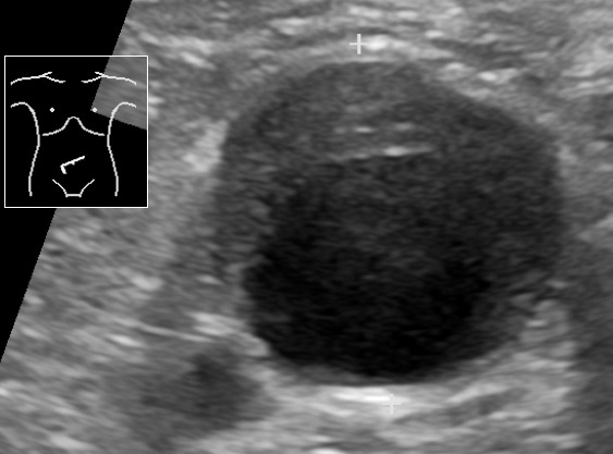
Ultrasonography of abdominal aortic aneurysm

Ultrasound showing an abdominal aortic aneurysm with a mural thrombus

Image: “Ultrasonography of abdominal aortic aneurysm with mural thrombus” by Mikael Häggström, M.D.. License: Public Domain

Management

Non-surgical management

  • Reduce cardiovascular risk:
    • Smoking Smoking Willful or deliberate act of inhaling and exhaling smoke from burning substances or agents held by hand. Interstitial Lung Diseases cessation (most effective nonsurgical intervention to reduce aneurysm-related complications and death)
    • Exercise:
      • Avoid heavy lifting.
      • Avoid activities that lead to Valsalva (↑ blood pressure).
    • Hypertension Hypertension Hypertension, or high blood pressure, is a common disease that manifests as elevated systemic arterial pressures. Hypertension is most often asymptomatic and is found incidentally as part of a routine physical examination or during triage for an unrelated medical encounter. Hypertension control:
      • Antihypertensives Antihypertensives The 1st-line medication classes for hypertension include thiazide-like diuretics, angiotensin-converting enzyme inhibitors (ACEis), angiotensin II receptor blockers (ARBs), and calcium channel blockers (CCBS). Contraindications, adverse effects, and drug-to-drug interactions are agent specific. Hypertension Drugs given to reach recommended blood pressure goals
      • Unlike TAA TAA Thoracic aortic aneurysm (TAA) is the abnormal dilation of a segment of the thoracic aorta, usually the ascending aorta. Most TAAs are due to degenerative aortic disorders, commonly in patients > 65 years of age. Most TAAs are asymptomatic (incidentally found in imaging) but could present with symptoms from its effects on surrounding structures. Thoracic Aortic Aneurysms, no specific medication recommended to limit Limit A value (e.g., pressure or time) that should not be exceeded and which is specified by the operator to protect the lung Invasive Mechanical Ventilation AAA expansion
    • Lipid control with statins Statins Statins are competitive inhibitors of HMG-CoA reductase in the liver. HMG-CoA reductase is the rate-limiting step in cholesterol synthesis. Inhibition results in lowered intrahepatocytic cholesterol formation, resulting in up-regulation of LDL receptors and, ultimately, lowering levels of serum LDL and triglycerides. Statins (target LDL < 70 mg/dL)
  • Avoid fluoroquinolones Fluoroquinolones Fluoroquinolones are a group of broad-spectrum, bactericidal antibiotics inhibiting bacterial DNA replication. Fluoroquinolones cover gram-negative, anaerobic, and atypical organisms, as well as some gram-positive and multidrug-resistant (MDR) organisms. Fluoroquinolones (may ↑ risk of dissection or rupture)
  • Surveillance Surveillance Developmental Milestones and Normal Growth: Asymptomatic AAA < 5.5 cm, periodic evaluation and aneurysm Aneurysm An aneurysm is a bulging, weakened area of a blood vessel that causes an abnormal widening of its diameter > 1.5 times the size of the native vessel. Aneurysms occur more often in arteries than in veins and are at risk of dissection and rupture, which can be life-threatening. Thoracic Aortic Aneurysms diameter surveillance Surveillance Developmental Milestones and Normal Growth
Table: Management of asymptomatic patients Patients Individuals participating in the health care system for the purpose of receiving therapeutic, diagnostic, or preventive procedures. Clinician–Patient Relationship by AAA size
AAA management of asymptomatic patients Patients Individuals participating in the health care system for the purpose of receiving therapeutic, diagnostic, or preventive procedures. Clinician–Patient Relationship (ultrasound) AAA size
Rescreen after 10 years ≥ 2.5 cm but < 3 cm
Imaging every 3 years 3–3.9 cm
Imaging every 12 months 4–4.9 cm
Imaging every 6 months 5–5.4 cm
Consider elective repair ≥ 5.5 cm

Consider repair if ≥ 5 cm in women; expansion of < 0.5 cm/6 months or > 1 cm/1 year; associated femoral, iliac, popliteal aneurysm Popliteal aneurysm Extremity and Visceral Aneurysms, or PAD requiring revascularization Revascularization Thromboangiitis Obliterans (Buerger’s Disease)

Surgical management

Indications for operative repair

  • AAA rupture: emergency repair
  • Elective repair: most effective way of preventing rupture and aneurysm-related death
    • Symptomatic AAA
    • Rapidly expanding (> 0.5 cm/6 months or > 1 cm/1 year)
    • Asymptomatic AAA:  when diameter ≥ 5.5 cm  (5 cm in women: higher rate of rupture)
    • AAA with coexisting iliac, femoral, or popliteal artery Popliteal Artery The continuation of the femoral artery coursing through the popliteal fossa; it divides into the anterior and posterior tibial arteries. Popliteal Fossa: Anatomy aneurysm Aneurysm An aneurysm is a bulging, weakened area of a blood vessel that causes an abnormal widening of its diameter > 1.5 times the size of the native vessel. Aneurysms occur more often in arteries than in veins and are at risk of dissection and rupture, which can be life-threatening. Thoracic Aortic Aneurysms
    • AAA associated with symptomatic peripheral arterial disease (PAD) undergoing revascularization Revascularization Thromboangiitis Obliterans (Buerger’s Disease)

Operative options

  • Open surgical repair: 
  • EVAR:
    • In the United States: 80% of AAA surgical repair 
    • Access through iliac or femoral arteries Arteries Arteries are tubular collections of cells that transport oxygenated blood and nutrients from the heart to the tissues of the body. The blood passes through the arteries in order of decreasing luminal diameter, starting in the largest artery (the aorta) and ending in the small arterioles. Arteries are classified into 3 types: large elastic arteries, medium muscular arteries, and small arteries and arterioles. Arteries: Histology and endograft placed within AAA lumen 
    • Requires anatomic suitability (site and structure of aneurysm Aneurysm An aneurysm is a bulging, weakened area of a blood vessel that causes an abnormal widening of its diameter > 1.5 times the size of the native vessel. Aneurysms occur more often in arteries than in veins and are at risk of dissection and rupture, which can be life-threatening. Thoracic Aortic Aneurysms and access vessels)
    • Decreased operative mortality Mortality All deaths reported in a given population. Measures of Health Status but higher rate of re-intervention
    • Surveillance Surveillance Developmental Milestones and Normal Growth:
      • CT angiography Angiography Radiography of blood vessels after injection of a contrast medium. Cardiac Surgery 1 month and 1 year post-operatively, then duplex ultrasonography Duplex ultrasonography Ultrasonography applying the doppler effect combined with real-time imaging. The real-time image is created by rapid movement of the ultrasound beam. A powerful advantage of this technique is the ability to estimate the velocity of flow from the doppler shift frequency. Hypercoagulable States/CT annually if with uncomplicated surgery 
      • Look for endoleak, sac enlargement, migration of stents, and device integrity

Differential Diagnosis

  • Ruptured viscus: a condition in which gastrointestinal wall integrity is lost with subsequent leakage of enteric contents Leakage of Enteric Contents Perforated Viscus into the peritoneal cavity Peritoneal Cavity The space enclosed by the peritoneum. It is divided into two portions, the greater sac and the lesser sac or omental bursa, which lies behind the stomach. The two sacs are connected by the foramen of winslow, or epiploic foramen. Peritoneum: Anatomy, resulting in peritonitis Peritonitis Inflammation of the peritoneum lining the abdominal cavity as the result of infectious, autoimmune, or chemical processes. Primary peritonitis is due to infection of the peritoneal cavity via hematogenous or lymphatic spread and without intra-abdominal source. Secondary peritonitis arises from the abdominal cavity itself through rupture or abscess of intra-abdominal organs. Penetrating Abdominal Injury. A ruptured viscus is life-threatening and requires surgical management.
  • Mesenteric ischemia Ischemia A hypoperfusion of the blood through an organ or tissue caused by a pathologic constriction or obstruction of its blood vessels, or an absence of blood circulation. Ischemic Cell Damage: a rare, life-threatening syndrome caused by inadequate blood flow Blood flow Blood flow refers to the movement of a certain volume of blood through the vasculature over a given unit of time (e.g., mL per minute). Vascular Resistance, Flow, and Mean Arterial Pressure through the mesenteric vessels resulting in ischemia Ischemia A hypoperfusion of the blood through an organ or tissue caused by a pathologic constriction or obstruction of its blood vessels, or an absence of blood circulation. Ischemic Cell Damage and gangrene Gangrene Death and putrefaction of tissue usually due to a loss of blood supply. Small Bowel Obstruction of the bowel wall. Mesenteric ischemia Ischemia A hypoperfusion of the blood through an organ or tissue caused by a pathologic constriction or obstruction of its blood vessels, or an absence of blood circulation. Ischemic Cell Damage can be acute or chronic. Acute mesenteric ischemia Acute Mesenteric Ischemia Mesenteric Ischemia is a surgical emergency Surgical Emergency Acute Abdomen, while the chronic condition requires risk factor modification, as it is related to vascular disease.
  • Strangulated hernia Hernia Protrusion of tissue, structure, or part of an organ through the bone, muscular tissue, or the membrane by which it is normally contained. Hernia may involve tissues such as the abdominal wall or the respiratory diaphragm. Hernias may be internal, external, congenital, or acquired. Abdominal Hernias: Hernias are protrusions of abdominal content ( peritoneum Peritoneum The peritoneum is a serous membrane lining the abdominopelvic cavity. This lining is formed by connective tissue and originates from the mesoderm. The membrane lines both the abdominal walls (as parietal peritoneum) and all of the visceral organs (as visceral peritoneum). Peritoneum: Anatomy, visceral fat, and/or viscera) through a congenital Congenital Chorioretinitis or acquired defect in the abdominal wall Abdominal wall The outer margins of the abdomen, extending from the osteocartilaginous thoracic cage to the pelvis. Though its major part is muscular, the abdominal wall consists of at least seven layers: the skin, subcutaneous fat, deep fascia; abdominal muscles, transversalis fascia, extraperitoneal fat, and the parietal peritoneum. Surgical Anatomy of the Abdomen. Strangulation Strangulation Inguinal Canal: Anatomy and Hernias involves the constriction of hernial contents leading to bowel ischemia Bowel ischemia Mesenteric ischemia is a rare, life-threatening condition caused by inadequate blood flow through the mesenteric vessels, which results in ischemia and necrosis of the intestinal wall. Mesenteric ischemia can be either acute or chronic. Mesenteric Ischemia and requires emergency surgery to avoid bowel loss, perforation Perforation A pathological hole in an organ, blood vessel or other soft part of the body, occurring in the absence of external force. Esophagitis, and sepsis Sepsis Systemic inflammatory response syndrome with a proven or suspected infectious etiology. When sepsis is associated with organ dysfunction distant from the site of infection, it is called severe sepsis. When sepsis is accompanied by hypotension despite adequate fluid infusion, it is called septic shock. Sepsis and Septic Shock.
  • Acute cholecystitis Acute cholecystitis Acute inflammation of the gallbladder wall. It is characterized by the presence of abdominal pain; fever; and leukocytosis. Gallstone obstruction of the cystic duct is present in approximately 90% of the cases. Cholecystitis: a condition characterized by inflammation Inflammation Inflammation is a complex set of responses to infection and injury involving leukocytes as the principal cellular mediators in the body’s defense against pathogenic organisms. Inflammation is also seen as a response to tissue injury in the process of wound healing. The 5 cardinal signs of inflammation are pain, heat, redness, swelling, and loss of function. Inflammation of the gallbladder Gallbladder The gallbladder is a pear-shaped sac, located directly beneath the liver, that sits on top of the superior part of the duodenum. The primary functions of the gallbladder include concentrating and storing up to 50 mL of bile. Gallbladder and Biliary Tract: Anatomy, most often due to obstruction of the cystic Cystic Fibrocystic Change duct by a gallstone. Management includes IV fluids IV fluids Intravenous fluids are one of the most common interventions administered in medicine to approximate physiologic bodily fluids. Intravenous fluids are divided into 2 categories: crystalloid and colloid solutions. Intravenous fluids have a wide variety of indications, including intravascular volume expansion, electrolyte manipulation, and maintenance fluids. Intravenous Fluids, pain Pain An unpleasant sensation induced by noxious stimuli which are detected by nerve endings of nociceptive neurons. Pain: Types and Pathways control, and IV antibiotics for secondary infection. Complicated cholecystitis Cholecystitis Cholecystitis is the inflammation of the gallbladder (GB) usually caused by the obstruction of the cystic duct (acute cholecystitis). Mechanical irritation by gallstones can also produce chronic GB inflammation. Cholecystitis is one of the most common complications of cholelithiasis but inflammation without gallstones can occur in a minority of patients. Cholecystitis and progressive symptoms are indications for emergency cholecystectomy Cholecystectomy Cholecystectomy is a surgical procedure performed with the goal of resecting and extracting the gallbladder. It is one of the most common abdominal surgeries performed in the Western world. Cholecystectomy is performed for symptomatic cholelithiasis, cholecystitis, gallbladder polyps > 0.5 cm, porcelain gallbladder, choledocholithiasis and gallstone pancreatitis, and rarely, for gallbladder cancer. Cholecystectomy.
  • Acute pancreatitis Pancreatitis Inflammation of the pancreas. Pancreatitis is classified as acute unless there are computed tomographic or endoscopic retrograde cholangiopancreatographic findings of chronic pancreatitis. The two most common forms of acute pancreatitis are alcoholic pancreatitis and gallstone pancreatitis. Acute Pancreatitis: an inflammation Inflammation Inflammation is a complex set of responses to infection and injury involving leukocytes as the principal cellular mediators in the body’s defense against pathogenic organisms. Inflammation is also seen as a response to tissue injury in the process of wound healing. The 5 cardinal signs of inflammation are pain, heat, redness, swelling, and loss of function. Inflammation of the pancreas Pancreas The pancreas lies mostly posterior to the stomach and extends across the posterior abdominal wall from the duodenum on the right to the spleen on the left. This organ has both exocrine and endocrine tissue. Pancreas: Anatomy that typically causes epigastric pain Epigastric pain Mallory-Weiss Syndrome (Mallory-Weiss Tear) that radiates to the back. This condition is often treated with aggressive fluid resuscitation Resuscitation The restoration to life or consciousness of one apparently dead. . Neonatal Respiratory Distress Syndrome, bowel rest, and pain Pain An unpleasant sensation induced by noxious stimuli which are detected by nerve endings of nociceptive neurons. Pain: Types and Pathways control. Surgery is indicated if the condition is associated with gallstones Gallstones Cholelithiasis (gallstones) is the presence of stones in the gallbladder. Most gallstones are cholesterol stones, while the rest are composed of bilirubin (pigment stones) and other mixed components. Patients are commonly asymptomatic but may present with biliary colic (intermittent pain in the right upper quadrant). Cholelithiasis.
  • Diverticular abscess Abscess Accumulation of purulent material in tissues, organs, or circumscribed spaces, usually associated with signs of infection. Chronic Granulomatous Disease: a group of various intestinal conditions characterized by abnormal outpouchings of the colonic mucosa (diverticula). Over time, these diverticula may accumulate intestinal content, become infected, swell, and develop into an abscess Abscess Accumulation of purulent material in tissues, organs, or circumscribed spaces, usually associated with signs of infection. Chronic Granulomatous Disease. Intravenous antibiotics are the recommended treatment, with percutaneous drainage Percutaneous Drainage Echinococcus/Echinococcosis needed for large abscess Abscess Accumulation of purulent material in tissues, organs, or circumscribed spaces, usually associated with signs of infection. Chronic Granulomatous Disease or failed medical treatment.

References

  1. Aortic aneurysm (n.d.). Cdc.gov. Retrieved 3 Sept 2020, from https://www.cdc.gov/heartdisease/aortic_aneurysm.htm
  2. Creager, M., Elmore, J. & Givens, J. (2020, Jul). Screening for abdominal aortic aneurysm. UpToDate. Retrieved 3 Sept 2020, from https://www.uptodate.com/contents/screening-for-abdominal-aortic-aneurysm
  3. Chung, J., Eidt, J., Mills, J., Creager, M., Collins, K. (2020, Mar). Epidemiology, risk factors, pathogenesis and natural history of abdominal aortic aneurysm. UpToDate. Retrieved 3 Sept 2020, from https://www.uptodate.com/contents/epidemiology-risk-factors-pathogenesis-and-natural-history-of-abdominal-aortic-aneurysm
  4. Dalman, R., Mell, M., Mills, J.; Eidt, J.,Clement, D., Creager, M., Collins, K. (Eds.).(2020, Jun).Management of asymptomatic aortic aneurysm. UpToDate. Retrieved 30 Aug 2020, from https://www.uptodate.com/contents/management-of-asymptomatic-abdominal-aortic-aneurysm
  5. Erbel R, Aboyans V, Boileau C, et al. (2014) ESC Guidelines on the diagnosis and treatment of aortic diseases: Document covering acute and chronic aortic diseases of the thoracic and abdominal aorta of the adult. The Task Force for the Diagnosis and Treatment of Aortic Diseases of the European Society of Cardiology (ESC). Eur Heart J .35:2873. Retrieved 30 Aug 2020, from https://www.escardio.org/Guidelines/Clinical-Practice-Guidelines/Aortic-Diseases
  6. Guoledge, J, Guo-Ping, S.; Norman, P., Fitridge, R., Thompson, M. (2011). Pathogenesis of aortic aneurysm in Mechanisms of Vascular disease: A Reference Book for Vascular Specialists. Adelaide (AU): University of Adelaide Press. Retrieved 29 Aug 2020, from https://www.ncbi.nlm.nih.gov/books/NBK534278/
  7. Mitchell, R., Halushka, M. (2020). In Kumar, V.; Abbas, A.; Aster, J.; Turner, J. (Eds.) Robbins and Cotran Pathologic Basis of Disease. (10th ed.,pp.485-525). Elsevier, Inc.
  8. Rahimi, S., Lopez Rowe, V. (2019, Jan). Abdominal aortic aneurysm. Medscape. Retrieved 28 Aug 2020, from https://emedicine.medscape.com/article/1979501-overview
  9. Ruddy, J., Jones, J.;Spinale, F.,Ikonomidis, J.(2008). Regional Heterogeneity within the Aorta: relevance to Aneurysm disease. J Thorac Cardiovasc Surg. 136(5): 1123–1130. Retrieved 5 Sept 2020, from https://www.ncbi.nlm.nih.gov/pmc/articles/PMC2679174/
  10. Sidloff, D., Stather, P., Dattani, N., Bown, M., Thompson, J., Sayers, R., Choke, E. (2013, Nov). Aneurysm Global epidemiology study. Circulation. Retrieved 3 Sept 2020, from https://www.ahajournals.org/doi/10.1161/CIRCULATIONAHA.113.005457
  11. Vu, K., Kaitoukov, Y., Morin-Roy, F., Kauffmann, C.,Giroux, F., Therasse, E., Soulez,G., Tang, A. (2014, Jun). Rupture signs on computed tomography, treatment, and outcome of abdominal aortic aneurysms. Insights Imaging.  5(3): 281–293. Retrieved 30 Aug 2020, from doi: 10.1007/s13244-014-0327-3

Create your free account or log in to continue reading!

Sign up now and get free access to Lecturio with concept pages, medical videos, and questions for your medical education.

User Reviews

Details