Achieve Mastery of Medical Concepts

Study for medical school and boards with Lecturio

Benign Breast Conditions

Benign Benign Fibroadenoma breast epithelial lesions are grouped histologically as nonproliferative, proliferative without atypia Atypia Fibrocystic Change, and atypical hyperplasia Hyperplasia An increase in the number of cells in a tissue or organ without tumor formation. It differs from hypertrophy, which is an increase in bulk without an increase in the number of cells. Cellular Adaptation. The classifications are based on subsequent cancer risk in either breast. The nonproliferative type carries no risk, while fibroadenoma Fibroadenoma Fibroadenomas are the most common benign tumor of the female breast and the most common breast tumor in adolescent and young women. The tumors are well-circumscribed, mobile, and unencapsulated, with a rubbery or firm consistency. Fibroadenoma, the most common benign Benign Fibroadenoma tumor Tumor Inflammation, is a proliferative breast lesion (i.e., has a slight increase in malignancy Malignancy Hemothorax risk). Because atypical hyperplasia Hyperplasia An increase in the number of cells in a tissue or organ without tumor formation. It differs from hypertrophy, which is an increase in bulk without an increase in the number of cells. Cellular Adaptation shares some features with breast carcinoma in situ Carcinoma in situ A lesion with cytological characteristics associated with invasive carcinoma but the tumor cells are confined to the epithelium of origin, without invasion of the basement membrane. Leukoplakia, future cancer potential is increased. Management ranges from frequent monitoring to surgical excision, depending on certain factors, including the inherent risk of the pathologic diagnosis. Other breast disorders without malignant possibility are associated with underlying infection or systemic disease, so treatment differs. Benign Benign Fibroadenoma breast diseases are common but present diversely. It is important to distinguish between them to determine the likelihood of cancer and the best course of treatment.

Last updated: Dec 5, 2022

Editorial responsibility: Stanley Oiseth, Lindsay Jones, Evelin Maza

Nonproliferative Epithelial Breast Lesions

Definition

Nonproliferative breast lesions are conditions generally not associated with an increased risk of breast cancer Breast cancer Breast cancer is a disease characterized by malignant transformation of the epithelial cells of the breast. Breast cancer is the most common form of cancer and 2nd most common cause of cancer-related death among women. Breast Cancer.
Simple breast cyst

  • Round or oval fluid-filled masses from the terminal duct lobular unit
  • Epidemiology:
    • Prevalence Prevalence The total number of cases of a given disease in a specified population at a designated time. It is differentiated from incidence, which refers to the number of new cases in the population at a given time. Measures of Disease Frequency: estimated to be 50%90%
    • Up to ⅓ of women aged 3050 have breast cysts Cysts Any fluid-filled closed cavity or sac that is lined by an epithelium. Cysts can be of normal, abnormal, non-neoplastic, or neoplastic tissues. Fibrocystic Change
  • Clinical presentation:
    • Felt as a palpable mass Mass Three-dimensional lesion that occupies a space within the breast Imaging of the Breast
    • Pain Pain An unpleasant sensation induced by noxious stimuli which are detected by nerve endings of nociceptive neurons. Pain: Types and Pathways noted with acute enlargement of cyst
  • Diagnosis:
  • Management:
    • No further intervention most of the time
    • Fine needle aspiration Fine Needle Aspiration Fibroadenoma
      • For signs of infection or inflammation Inflammation Inflammation is a complex set of responses to infection and injury involving leukocytes as the principal cellular mediators in the body’s defense against pathogenic organisms. Inflammation is also seen as a response to tissue injury in the process of wound healing. The 5 cardinal signs of inflammation are pain, heat, redness, swelling, and loss of function. Inflammation 
      • Pain Pain An unpleasant sensation induced by noxious stimuli which are detected by nerve endings of nociceptive neurons. Pain: Types and Pathways from increased size

Mild hyperplasia Hyperplasia An increase in the number of cells in a tissue or organ without tumor formation. It differs from hypertrophy, which is an increase in bulk without an increase in the number of cells. Cellular Adaptation of the usual type

  • Pathology:
    • Cells look very close to normal. 
    • An increase in the number of epithelial cells within a duct
    • Cell depth is more than 2 but not more than 4 cells.
  • Clinical presentation: found on biopsy Biopsy Removal and pathologic examination of specimens from the living body. Ewing Sarcoma or surgical excision
  • Management: no treatment needed

Papillary apocrine change

  • Pathology:
    • Proliferation of ductal epithelial cells with apocrine features
    • May be simple, complex, or highly complex lesions 
  • Clinical presentation: found on histological examination ( biopsy Biopsy Removal and pathologic examination of specimens from the living body. Ewing Sarcoma or surgical excision)
Breast usg

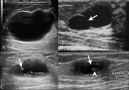
Breast US shows a simple cyst (A) presenting as an anechoic lesion with posterior enhancement and a cyst (B) with a septum (arrow). Breast US (C) in a 26-year-old woman with a painless palpable lump in her left breast shows a complex cyst with an eccentrically placed echogenic focus (arrow) representing the scolex of a cysticercus granuloma. The patient also had a similar swelling in the right upper arm, a US (D) of which revealed a cyst (arrow) with an echogenic scolex (arrowhead), within.

Image: “Breast USG” by Advanced Radiology Centre, Mumbai, India. License: CC BY 2.0

Proliferative Epithelial Lesions without Atypia

Definition

In proliferative breast lesions without atypia Atypia Fibrocystic Change, the development of breast cancer Breast cancer Breast cancer is a disease characterized by malignant transformation of the epithelial cells of the breast. Breast cancer is the most common form of cancer and 2nd most common cause of cancer-related death among women. Breast Cancer is 1.5–2 times the risk of the general population.
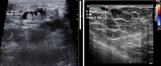
Intraductal papilloma Papilloma A circumscribed benign epithelial tumor projecting from the surrounding surface; more precisely, a benign epithelial neoplasm consisting of villous or arborescent outgrowths of fibrovascular stroma covered by neoplastic cells. Cowden Syndrome

  • Benign Benign Fibroadenoma mass Mass Three-dimensional lesion that occupies a space within the breast Imaging of the Breast with a papillary configuration of breast stroma and epithelium Epithelium The epithelium is a complex of specialized cellular organizations arranged into sheets and lining cavities and covering the surfaces of the body. The cells exhibit polarity, having an apical and a basal pole. Structures important for the epithelial integrity and function involve the basement membrane, the semipermeable sheet on which the cells rest, and interdigitations, as well as cellular junctions. Surface Epithelium: Histology within a breast duct
  • Epidemiology:
    • Peak incidence Incidence The number of new cases of a given disease during a given period in a specified population. It also is used for the rate at which new events occur in a defined population. It is differentiated from prevalence, which refers to all cases in the population at a given time. Measures of Disease Frequency: 3050 years of age
    • < 10% of benign Benign Fibroadenoma breast lesions
    • High-risk precursor lesion with the following predisposing factors:
      • Contraceptive use
      • Hormone replacement therapy Hormone Replacement Therapy Hormone replacement therapy (HRT) is used to treat symptoms associated with female menopause and in combination to suppress ovulation. Risks and side effects include uterine bleeding, predisposition to cancer, breast tenderness, hyperpigmentation, migraine headaches, hypertension, bloating, and mood changes. Noncontraceptive Estrogen and Progestins
      • Family history Family History Adult Health Maintenance of breast cancer Breast cancer Breast cancer is a disease characterized by malignant transformation of the epithelial cells of the breast. Breast cancer is the most common form of cancer and 2nd most common cause of cancer-related death among women. Breast Cancer
  • Clinical presentation: depends on the size and location
    • Central type:
    • Peripheral type: (10% of cases)
      • Often appears as multiple papillomas and affects younger premenopausal women 
      • Found on the outer areas of the breast (growth in terminal ducts)
      • Usually non-palpable and less likely to have discharge
      • Appears as mammographic calcifications
      • Increased risk for breast cancer Breast cancer Breast cancer is a disease characterized by malignant transformation of the epithelial cells of the breast. Breast cancer is the most common form of cancer and 2nd most common cause of cancer-related death among women. Breast Cancer
  • Diagnosis:
    • Mammography Mammography Radiographic examination of the breast. Breast Cancer Screening:
      • Can be occult
      • When seen, oval/round mass Mass Three-dimensional lesion that occupies a space within the breast Imaging of the Breast; margins can be ill-defined
      • May have calcifications
    • Core needle biopsy Core Needle Biopsy Fibrocystic Change (CNB) (guided by ductogram or US):
      • Preferred over fine needle biopsy Biopsy Removal and pathologic examination of specimens from the living body. Ewing Sarcoma (risk of inadequate tissue sample)
      • Shows papillary cells with a central branching fibrovascular core
  • Management: 
    • Surgical excision/ lumpectomy Lumpectomy Fat Necrosis of the Breast with complete removal: due to risk of atypical ductal hyperplasia Hyperplasia An increase in the number of cells in a tissue or organ without tumor formation. It differs from hypertrophy, which is an increase in bulk without an increase in the number of cells. Cellular Adaptation or ductal carcinoma in situ Carcinoma in situ A lesion with cytological characteristics associated with invasive carcinoma but the tumor cells are confined to the epithelium of origin, without invasion of the basement membrane. Leukoplakia ( DCIS DCIS Proliferation of cytologically malignant cells within the mammary ductal system, with no invasion of the surrounding stroma Breast Cancer)
    • Annual breast exam and mammogram Mammogram Fibrocystic Change

Simple fibroadenoma Fibroadenoma Fibroadenomas are the most common benign tumor of the female breast and the most common breast tumor in adolescent and young women. The tumors are well-circumscribed, mobile, and unencapsulated, with a rubbery or firm consistency. Fibroadenoma

  • A benign Benign Fibroadenoma solid breast mass Mass Three-dimensional lesion that occupies a space within the breast Imaging of the Breast composed of fibrous Fibrous Fibrocystic Change and glandular tissue
  • Epidemiology:
    • Most common benign Benign Fibroadenoma tumor Tumor Inflammation of the breast
    • Peak incidence Incidence The number of new cases of a given disease during a given period in a specified population. It also is used for the rate at which new events occur in a defined population. It is differentiated from prevalence, which refers to all cases in the population at a given time. Measures of Disease Frequency: 1535 years of age
    • For majority, no potential for cancer
    • Risk factors of subsequent cancer:
      • Proliferative histology
      • Complex mass Mass Three-dimensional lesion that occupies a space within the breast Imaging of the Breast 
      • Family history Family History Adult Health Maintenance of breast cancer Breast cancer Breast cancer is a disease characterized by malignant transformation of the epithelial cells of the breast. Breast cancer is the most common form of cancer and 2nd most common cause of cancer-related death among women. Breast Cancer
  • Clinical presentation:
    • Small, well-defined, spherical, mobile mass Mass Three-dimensional lesion that occupies a space within the breast Imaging of the Breast 
    • Rubbery or firm consistency Consistency Dermatologic Examination, non-tender
    • Size: usually ≤ 3 cm in diameter, solitary
    • Location: often in the upper outer quadrant
    • Mass Mass Three-dimensional lesion that occupies a space within the breast Imaging of the Breast size and tenderness influenced by estrogen Estrogen Compounds that interact with estrogen receptors in target tissues to bring about the effects similar to those of estradiol. Estrogens stimulate the female reproductive organs, and the development of secondary female sex characteristics. Estrogenic chemicals include natural, synthetic, steroidal, or non-steroidal compounds. Ovaries: Anatomy levels:
      • Increased size: pregnancy Pregnancy The status during which female mammals carry their developing young (embryos or fetuses) in utero before birth, beginning from fertilization to birth. Pregnancy: Diagnosis, Physiology, and Care, lactation Lactation The processes of milk secretion by the maternal mammary glands after parturition. The proliferation of the mammary glandular tissue, milk synthesis, and milk expulsion or let down are regulated by the interactions of several hormones including estradiol; progesterone; prolactin; and oxytocin. Breastfeeding, pre-menstruation, and oral contraceptives
      • Regresses after menopause Menopause Menopause is a physiologic process in women characterized by the permanent cessation of menstruation that occurs after the loss of ovarian activity. Menopause can only be diagnosed retrospectively, after 12 months without menstrual bleeding. Menopause
  • Diagnosis:
    • US: well-defined solid mass Mass Three-dimensional lesion that occupies a space within the breast Imaging of the Breast, regular Regular Insulin boundaries, and a weak echo signal
    • Mammogram Mammogram Fibrocystic Change:
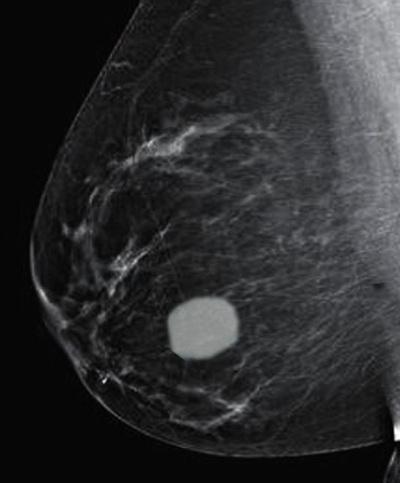
      • Well-circumscribed oval mass Mass Three-dimensional lesion that occupies a space within the breast Imaging of the Breast, may have lobulations
      • Popcorn-like calcifications in involuting fibroadenomas
    • Biopsy Biopsy Removal and pathologic examination of specimens from the living body. Ewing Sarcoma:
      • Indications:
        • > 5 cm or increasing size
        • Irregular borders 
        • Considerable pain Pain An unpleasant sensation induced by noxious stimuli which are detected by nerve endings of nociceptive neurons. Pain: Types and Pathways
      • Cytology: uniformly distributed epithelial cells (honeycomb or antler-like pattern)
      • Microscopy: stroma and epithelial cells either in pericanalicular or intracanalicular pattern
    • MRI: may have dark internal septations
  • Management:
    • Fibroadenoma Fibroadenoma Fibroadenomas are the most common benign tumor of the female breast and the most common breast tumor in adolescent and young women. The tumors are well-circumscribed, mobile, and unencapsulated, with a rubbery or firm consistency. Fibroadenoma with benign Benign Fibroadenoma features: observation, breast exam, and annual mammogram Mammogram Fibrocystic Change
    • Cryoablation Cryoablation Fibroadenoma: an option for small but symptomatic fibroadenomas
      • CNB done first to confirm diagnosis
      • Criteria:
        • For fibroadenomas < 4 cm in size, < 3 in number
        • No bleeding tendency and no local skin Skin The skin, also referred to as the integumentary system, is the largest organ of the body. The skin is primarily composed of the epidermis (outer layer) and dermis (deep layer). The epidermis is primarily composed of keratinocytes that undergo rapid turnover, while the dermis contains dense layers of connective tissue. Skin: Structure and Functions infection 
    • Surgical excision for:
      • Adolescent-onset fibroadenoma Fibroadenoma Fibroadenomas are the most common benign tumor of the female breast and the most common breast tumor in adolescent and young women. The tumors are well-circumscribed, mobile, and unencapsulated, with a rubbery or firm consistency. Fibroadenoma persisting through adulthood (risk of breast cancer Breast cancer Breast cancer is a disease characterized by malignant transformation of the epithelial cells of the breast. Breast cancer is the most common form of cancer and 2nd most common cause of cancer-related death among women. Breast Cancer increases)
      • Increase in size or with atypical features or symptoms

Related videos

Radial scars

Sclerosing adenosis Sclerosing Adenosis Fibroadenoma

Usual ductal hyperplasia Hyperplasia An increase in the number of cells in a tissue or organ without tumor formation. It differs from hypertrophy, which is an increase in bulk without an increase in the number of cells. Cellular Adaptation

Ductal hyperplasia

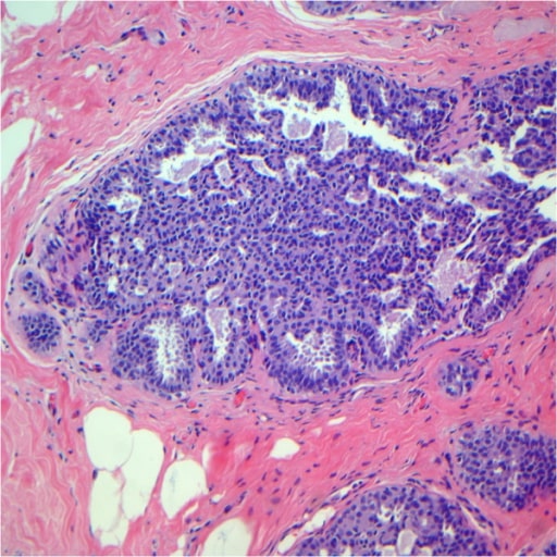
Usual ductal hyperplasia after review. Note the epithelial cells displaying a haphazard orientation and the presence of slit-like secondary lumina peripherally located.

Image: “Usual ductal hyperplasia” by Breast Pathology Laboratory, School of Medicine, Federal University of Minas Gerais (UFMG), Av, Professor Alfredo Balena, 190, Belo Horizonte, Minas Gerais 30130-100, Brazil. License: CC BY 2.0
Benign Epithelial Breast Lesions with Atypical Hyperplasia

Atypical ductal hyperplasia Hyperplasia An increase in the number of cells in a tissue or organ without tumor formation. It differs from hypertrophy, which is an increase in bulk without an increase in the number of cells. Cellular Adaptation (ADH)

  • Found in 5%20% of breast biopsies
  • Moderate increase in breast cancer Breast cancer Breast cancer is a disease characterized by malignant transformation of the epithelial cells of the breast. Breast cancer is the most common form of cancer and 2nd most common cause of cancer-related death among women. Breast Cancer risk
  • Clinical presentation: too small for palpation Palpation Application of fingers with light pressure to the surface of the body to determine consistency of parts beneath in physical diagnosis; includes palpation for determining the outlines of organs. Dermatologic Examination, discovered incidentally on biopsy Biopsy Removal and pathologic examination of specimens from the living body. Ewing Sarcoma (for mammographic findings such as calcifications)
  • Diagnosis: 
    • CNB
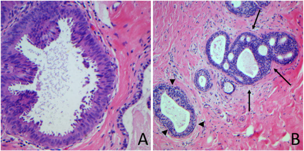
    • Pathology: proliferation of monomorphic epithelial cells in the breast duct
    • Shares features of low-grade DCIS DCIS Proliferation of cytologically malignant cells within the mammary ductal system, with no invasion of the surrounding stroma Breast Cancer without meeting full criteria
  • Management: 
    • Surgical excision: standard of care Standard of care The minimum acceptable patient care, based on statutes, court decisions, policies, or professional guidelines. Malpractice (ADH is premalignant) 
    • Breast exam every 612 months and annual mammogram Mammogram Fibrocystic Change
    • Avoid hormone replacement therapy Hormone Replacement Therapy Hormone replacement therapy (HRT) is used to treat symptoms associated with female menopause and in combination to suppress ovulation. Risks and side effects include uterine bleeding, predisposition to cancer, breast tenderness, hyperpigmentation, migraine headaches, hypertension, bloating, and mood changes. Noncontraceptive Estrogen and Progestins, oral contraceptives
    • Lifestyle and dietary changes
Atypical ductal hyperplasia

Atypical ductal hyperplasia (arrows)

Image: “Atypical ductal hyperplasia” by Breast Pathology Laboratory, School of Medicine, Federal University of Minas Gerais (UFMG), Av, Professor Alfredo Balena, 190, Belo Horizonte, Minas Gerais 30130-100, Brazil. License: CC BY 2.0
Atypical lobular hyperplasia Hyperplasia An increase in the number of cells in a tissue or organ without tumor formation. It differs from hypertrophy, which is an increase in bulk without an increase in the number of cells. Cellular Adaptation (ALH)

  • Increased risk of developing breast cancer Breast cancer Breast cancer is a disease characterized by malignant transformation of the epithelial cells of the breast. Breast cancer is the most common form of cancer and 2nd most common cause of cancer-related death among women. Breast Cancer in either ipsilateral or contralateral breast
  • Clinical presentation: discovered incidentally on biopsy Biopsy Removal and pathologic examination of specimens from the living body. Ewing Sarcoma (for mammographic findings such as calcifications)
  • Diagnosis:
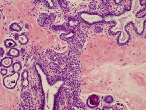
    • Pathology: Within the acini of terminal duct lobular unit, there is over-proliferation of dyshesive epithelial cells.
    • Shares features of lobular carcinoma in situ Carcinoma in situ A lesion with cytological characteristics associated with invasive carcinoma but the tumor cells are confined to the epithelium of origin, without invasion of the basement membrane. Leukoplakia or invasive lobular carcinoma without meeting full criteria
  • Management:
    • Surgical excision is generally appropriate but not routine.
    • Excision for high-risk patients Patients Individuals participating in the health care system for the purpose of receiving therapeutic, diagnostic, or preventive procedures. Clinician–Patient Relationship (family/personal history of breast cancer Breast cancer Breast cancer is a disease characterized by malignant transformation of the epithelial cells of the breast. Breast cancer is the most common form of cancer and 2nd most common cause of cancer-related death among women. Breast Cancer, BRCA1 or BRCA2 mutation Mutation Genetic mutations are errors in DNA that can cause protein misfolding and dysfunction. There are various types of mutations, including chromosomal, point, frameshift, and expansion mutations. Types of Mutations)
    • In carefully selected lower-risk patients Patients Individuals participating in the health care system for the purpose of receiving therapeutic, diagnostic, or preventive procedures. Clinician–Patient Relationship:
      • Estrogen Estrogen Compounds that interact with estrogen receptors in target tissues to bring about the effects similar to those of estradiol. Estrogens stimulate the female reproductive organs, and the development of secondary female sex characteristics. Estrogenic chemicals include natural, synthetic, steroidal, or non-steroidal compounds. Ovaries: Anatomy receptor Receptor Receptors are proteins located either on the surface of or within a cell that can bind to signaling molecules known as ligands (e.g., hormones) and cause some type of response within the cell. Receptors modulators
      • Increased mammogram Mammogram Fibrocystic Change and follow-up frequency 
    • Risk-reduction measures:
      • No hormone replacement therapy Hormone Replacement Therapy Hormone replacement therapy (HRT) is used to treat symptoms associated with female menopause and in combination to suppress ovulation. Risks and side effects include uterine bleeding, predisposition to cancer, breast tenderness, hyperpigmentation, migraine headaches, hypertension, bloating, and mood changes. Noncontraceptive Estrogen and Progestins, oral contraceptives
      • Lifestyle and dietary changes
Atypical lobular hyperplasia

Photomicrograph of atypical lobular hyperplasia associated with columnar cell changes and incidental to targeted microcalcifications

Image: “Atypical lobular hyperplasia” by Department of Pathology, The University of Texas MD Anderson Cancer Center, Houston, Texas. License: CC BY 3.0
Flat Epithelial Atypia

  • A distinct lesion from ADH and ALH 
  • Flat growth pattern (does not meet architectural criteria of ADH)
  • High-risk or atypical breast lesion but risk remains undefined
  • Clinical presentation: discovered on biopsies done for mammographic calcifications
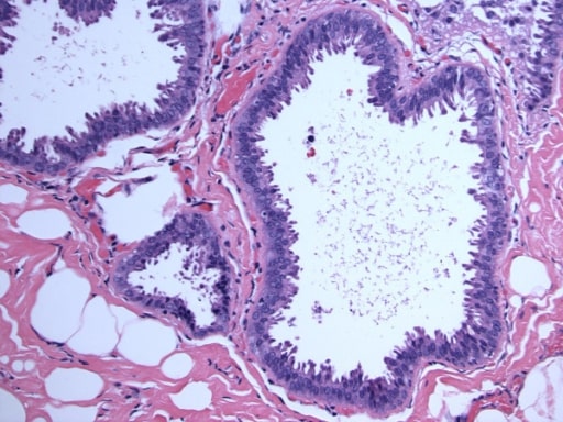
  • Diagnosis: pathology characterized by columnar changes with atypia Atypia Fibrocystic Change
  • Management:
    • Surgical consultation with risk assessment Risk assessment The qualitative or quantitative estimation of the likelihood of adverse effects that may result from exposure to specified health hazards or from the absence of beneficial influences. Preoperative Care
    • Observation is an option along with frequent exam and mammogram Mammogram Fibrocystic Change.
Flat epithelial atypia

Columnar cell lesions with atypia (flat epithelial atypia)

Image: “Flat epithelial atypia” by Department of Pathology, Wilford Hall Medical Center, Lackland AFB, TX, USA. License: CC BY 2.0
Breast Abscess

  • Epidemiology: usually associated with mastitis Mastitis Mastitis is inflammation of the breast tissue with or without infection. The most common form of mastitis is associated with lactation in the first few weeks after birth. Non-lactational mastitis includes periductal mastitis and idiopathic granulomatous mastitis (IGM). Mastitis
    • Known risk factors (in lactating women):
    • Known risk factors (in non-lactating women):
      • African American
      • Obesity Obesity Obesity is a condition associated with excess body weight, specifically with the deposition of excessive adipose tissue. Obesity is considered a global epidemic. Major influences come from the western diet and sedentary lifestyles, but the exact mechanisms likely include a mixture of genetic and environmental factors. Obesity 
      • Diabetes Diabetes Diabetes mellitus (DM) is a metabolic disease characterized by hyperglycemia and dysfunction of the regulation of glucose metabolism by insulin. Type 1 DM is diagnosed mostly in children and young adults as the result of autoimmune destruction of β cells in the pancreas and the resulting lack of insulin. Type 2 DM has a significant association with obesity and is characterized by insulin resistance. Diabetes Mellitus 
      • Smoking Smoking Willful or deliberate act of inhaling and exhaling smoke from burning substances or agents held by hand. Interstitial Lung Diseases
  • Clinical presentation:
    • Unilateral and fluctuant Fluctuant Dermatologic Examination mass Mass Three-dimensional lesion that occupies a space within the breast Imaging of the Breast
    • Painful, erythematous, and edematous breast 
    • Possible purulent discharge Purulent Discharge Dacryocystitis from the nipple Nipple The conic organs which usually give outlet to milk from the mammary glands. Examination of the Breast
    • Fever Fever Fever is defined as a measured body temperature of at least 38°C (100.4°F). Fever is caused by circulating endogenous and/or exogenous pyrogens that increase levels of prostaglandin E2 in the hypothalamus. Fever is commonly associated with chills, rigors, sweating, and flushing of the skin. Fever
  • Diagnosis:
  • Management:
    • Incision and drainage Incision And Drainage Chalazion especially:
      • If with skin Skin The skin, also referred to as the integumentary system, is the largest organ of the body. The skin is primarily composed of the epidermis (outer layer) and dermis (deep layer). The epidermis is primarily composed of keratinocytes that undergo rapid turnover, while the dermis contains dense layers of connective tissue. Skin: Structure and Functions ischemia Ischemia A hypoperfusion of the blood through an organ or tissue caused by a pathologic constriction or obstruction of its blood vessels, or an absence of blood circulation. Ischemic Cell Damage or pressure necrosis Necrosis The death of cells in an organ or tissue due to disease, injury or failure of the blood supply. Ischemic Cell Damage
      • If needle aspiration Needle aspiration Using fine needles (finer than 22-gauge) to remove tissue or fluid specimens from the living body for examination in the pathology laboratory and for disease diagnosis. Peritonsillar Abscess and/or antibiotics fail
    • Needle aspiration Needle aspiration Using fine needles (finer than 22-gauge) to remove tissue or fluid specimens from the living body for examination in the pathology laboratory and for disease diagnosis. Peritonsillar Abscess:
      •  Can be an initial treatment if overlying skin Skin The skin, also referred to as the integumentary system, is the largest organ of the body. The skin is primarily composed of the epidermis (outer layer) and dermis (deep layer). The epidermis is primarily composed of keratinocytes that undergo rapid turnover, while the dermis contains dense layers of connective tissue. Skin: Structure and Functions is not ischemic or abscess Abscess Accumulation of purulent material in tissues, organs, or circumscribed spaces, usually associated with signs of infection. Chronic Granulomatous Disease(es) < 3 cm
      • Repeated every 23 days until there is no collection
    • Antibiotics that target most common causative agent, Staphylococcus aureus Staphylococcus aureus Potentially pathogenic bacteria found in nasal membranes, skin, hair follicles, and perineum of warm-blooded animals. They may cause a wide range of infections and intoxications. Brain Abscess: cephalexin, dicloxacillin Dicloxacillin One of the penicillins which is resistant to penicillinase. Penicillins, or amoxicillin-clavulanate
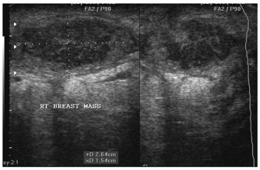
Breast abscess

Sonogram of a 22-year-old woman showing a right breast abscess. Note the oval shape of the abscess, which measures 2.64 cm by 1.54 cm before aspiration under ultrasound guidance.

Image: “Breast abscess” by Department of Surgery, Weil Bugando University College of Health Sciences, Mwanza, Tanzania. License: CC BY 2.0
Miscellaneous Breast Lesions

Diabetic mastopathy

  • Lymphocytic mastopathy or lymphocytic mastitis Mastitis Mastitis is inflammation of the breast tissue with or without infection. The most common form of mastitis is associated with lactation in the first few weeks after birth. Non-lactational mastitis includes periductal mastitis and idiopathic granulomatous mastitis (IGM). Mastitis
  • May be due to secondary autoimmune reaction from effects of hyperglycemia Hyperglycemia Abnormally high blood glucose level. Diabetes Mellitus on connective tissue Connective tissue Connective tissues originate from embryonic mesenchyme and are present throughout the body except inside the brain and spinal cord. The main function of connective tissues is to provide structural support to organs. Connective tissues consist of cells and an extracellular matrix. Connective Tissue: Histology
  • Seen in up to 13% of patients Patients Individuals participating in the health care system for the purpose of receiving therapeutic, diagnostic, or preventive procedures. Clinician–Patient Relationship with diabetes Diabetes Diabetes mellitus (DM) is a metabolic disease characterized by hyperglycemia and dysfunction of the regulation of glucose metabolism by insulin. Type 1 DM is diagnosed mostly in children and young adults as the result of autoimmune destruction of β cells in the pancreas and the resulting lack of insulin. Type 2 DM has a significant association with obesity and is characterized by insulin resistance. Diabetes Mellitus mellitus type 1 Type 1 Spinal Muscular Atrophy
  • Clinical presentation: painless mass Mass Three-dimensional lesion that occupies a space within the breast Imaging of the Breast, seen in long-standing diabetes Diabetes Diabetes mellitus (DM) is a metabolic disease characterized by hyperglycemia and dysfunction of the regulation of glucose metabolism by insulin. Type 1 DM is diagnosed mostly in children and young adults as the result of autoimmune destruction of β cells in the pancreas and the resulting lack of insulin. Type 2 DM has a significant association with obesity and is characterized by insulin resistance. Diabetes Mellitus mellitus type 1 Type 1 Spinal Muscular Atrophy
  • Diagnosis:
  • Management:
    • Excision not needed as there is no increased risk for breast cancer Breast cancer Breast cancer is a disease characterized by malignant transformation of the epithelial cells of the breast. Breast cancer is the most common form of cancer and 2nd most common cause of cancer-related death among women. Breast Cancer
    • Known to recur after surgical removal
    • Annual mammogram Mammogram Fibrocystic Change

Sarcoidosis Sarcoidosis Sarcoidosis is a multisystem inflammatory disease that causes noncaseating granulomas. The exact etiology is unknown. Sarcoidosis usually affects the lungs and thoracic lymph nodes, but it can also affect almost every system in the body, including the skin, heart, and eyes, most commonly. Sarcoidosis

  • Seen in patients Patients Individuals participating in the health care system for the purpose of receiving therapeutic, diagnostic, or preventive procedures. Clinician–Patient Relationship with systemic sarcoidosis Sarcoidosis Sarcoidosis is a multisystem inflammatory disease that causes noncaseating granulomas. The exact etiology is unknown. Sarcoidosis usually affects the lungs and thoracic lymph nodes, but it can also affect almost every system in the body, including the skin, heart, and eyes, most commonly. Sarcoidosis but mammary involvement can be the sole manifestation
  • Clinical presentation: firm breast masses
  • Diagnosis:
  • Management: 
    • Glucocorticoids Glucocorticoids Glucocorticoids are a class within the corticosteroid family. Glucocorticoids are chemically and functionally similar to endogenous cortisol. There are a wide array of indications, which primarily benefit from the antiinflammatory and immunosuppressive effects of this class of drugs. Glucocorticoids
    • Surgical excision an option

Galactocele

  • A retention cyst within the mammary gland Mammary gland Glandular tissue in the breast of human that is under the influence of hormones such as estrogens; progestins; and prolactin. In women, after parturition, the mammary glands secrete milk for the nourishment of the young. Breasts: Anatomy containing milk 
  • An obstruction of a lactiferous duct → accumulation of epithelial cells and milk → distention of the duct → cyst formation
  • Epidemiology: most common benign Benign Fibroadenoma breast condition in lactating women
  • Clinical presentation:
    • Palpable, firm mass Mass Three-dimensional lesion that occupies a space within the breast Imaging of the Breast in the subareolar region
    • Presents with no fever Fever Fever is defined as a measured body temperature of at least 38°C (100.4°F). Fever is caused by circulating endogenous and/or exogenous pyrogens that increase levels of prostaglandin E2 in the hypothalamus. Fever is commonly associated with chills, rigors, sweating, and flushing of the skin. Fever or pain Pain An unpleasant sensation induced by noxious stimuli which are detected by nerve endings of nociceptive neurons. Pain: Types and Pathways ( pain Pain An unpleasant sensation induced by noxious stimuli which are detected by nerve endings of nociceptive neurons. Pain: Types and Pathways suggests secondary bacterial infection)
    • Unilateral
  • Diagnosis:
  • Management: 
    • Most cases resolve spontaneously.
    • Increased breastfeeding Breastfeeding Breastfeeding is often the primary source of nutrition for the newborn. During pregnancy, hormonal stimulation causes the number and size of mammary glands in the breast to significantly increase. After delivery, prolactin stimulates milk production, while oxytocin stimulates milk expulsion through the lactiferous ducts, where it is sucked out through the nipple by the infant. Breastfeeding, warm compresses Warm Compresses Chalazion, and massage 
    • Repeated needle aspiration Needle aspiration Using fine needles (finer than 22-gauge) to remove tissue or fluid specimens from the living body for examination in the pathology laboratory and for disease diagnosis. Peritonsillar Abscess or surgical excision: for symptomatic cysts Cysts Any fluid-filled closed cavity or sac that is lined by an epithelium. Cysts can be of normal, abnormal, non-neoplastic, or neoplastic tissues. Fibrocystic Change
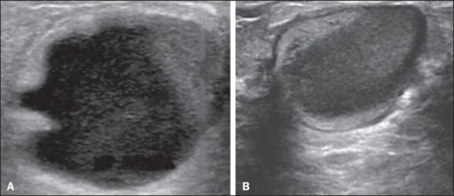
Galactocele

Galactocele:
A: Lesion parallel to the skin with well-defined borders showing anechoic (cystic) and echogenic (solid) components, with discrete posterior acoustic enhancement and well-defined borders.
B: Predominantly hypoechoic lesion parallel to the skin with well-defined borders, peripheral areas of hyperechogenicity, and posterior acoustic enhancement.
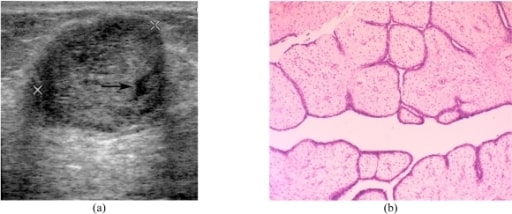
Image: “ Galactocele” by US National Library of Medicine. License: CC BY 4.0

Phyllodes tumor Tumor Inflammation

  • Cystosarcoma phyllodes
  • Fibroepithelial tumor Tumor Inflammation similar to fibroadenomas, characterized by rapid growth
  • Epidemiology:
    • Most common in women between 40 and 50 years of age
    • Account for < 1% of all breast neoplasms Neoplasms New abnormal growth of tissue. Malignant neoplasms show a greater degree of anaplasia and have the properties of invasion and metastasis, compared to benign neoplasms. Benign Bone Tumors
    • Although usually benign Benign Fibroadenoma, some may become malignant (25% of cases)
  • Clinical presentation:
    • Painless, multinodular, firm, rapidly growing mass Mass Three-dimensional lesion that occupies a space within the breast Imaging of the Breast 
    • Usually 47 cm in diameter
    • About 20% nonpalpable, identified on mammography Mammography Radiographic examination of the breast. Breast Cancer Screening
  • Diagnosis:
    • Mammography Mammography Radiographic examination of the breast. Breast Cancer Screening: lobulated mass Mass Three-dimensional lesion that occupies a space within the breast Imaging of the Breast, resembles fibroadenoma Fibroadenoma Fibroadenomas are the most common benign tumor of the female breast and the most common breast tumor in adolescent and young women. The tumors are well-circumscribed, mobile, and unencapsulated, with a rubbery or firm consistency. Fibroadenoma
    • Ultrasonography: well-defined, hypoechoic Hypoechoic A structure that produces a low-amplitude echo (darker grays) Ultrasound (Sonography) mass Mass Three-dimensional lesion that occupies a space within the breast Imaging of the Breast (like fibroadenomas, without microcalcifications)
    • MRI: well-circumscribed with irregular walls, high signal intensity on T1, and low signal intensity on T2 
    • CNB:
      • Papillary projection (phyllodes/“leaf-like”) of proliferating epithelium-lined stroma 
      • Varying degrees of atypia Atypia Fibrocystic Change and hyperplasia Hyperplasia An increase in the number of cells in a tissue or organ without tumor formation. It differs from hypertrophy, which is an increase in bulk without an increase in the number of cells. Cellular Adaptation (depends on benign Benign Fibroadenoma or malignant nature)
  • Management: 
    • Surgical resection with wide margins (due to high recurrence rate)
    • Breast exam and mammogram Mammogram Fibrocystic Change every 6 months for 2 years, then annually
Phyllodes tumor

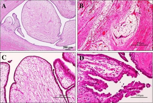
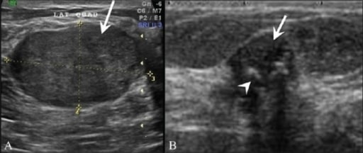
Phyllodes tumor of the breast

(a) Transverse US image shows a circumscribed heterogenous echo with a small cystic space (arrow) and a slight posterior acoustic enhancement.
(b) Photomicrograph shows leaf-like processes containing cellular stroma lined with benign ductal epithelial cells projecting into the cystic space (haematoxylin & eosin stain; x100).

Image: “Benign phyllodes tumour in a 35-year-old woman” by Department of Radiology, Chiang Mai University, Chiang Mai, Thailand. License: CC BY 2.5

Giant juvenile fibroadenoma Fibroadenoma Fibroadenomas are the most common benign tumor of the female breast and the most common breast tumor in adolescent and young women. The tumors are well-circumscribed, mobile, and unencapsulated, with a rubbery or firm consistency. Fibroadenoma

  • Approximately 0.5%2% of all fibroadenomas
  • Usually occurs in African American adolescents
  • Clinical presentation:
    • Rapid increase in size; unilateral tumors > 5 cm in diameter
    • May cause skin Skin The skin, also referred to as the integumentary system, is the largest organ of the body. The skin is primarily composed of the epidermis (outer layer) and dermis (deep layer). The epidermis is primarily composed of keratinocytes that undergo rapid turnover, while the dermis contains dense layers of connective tissue. Skin: Structure and Functions ulceration Ulceration Corneal Abrasions, Erosion, and Ulcers and venous engorgement Engorgement Mastitis
  • Management: surgical excision (as they are not easily distinguished from phyllodes tumors on examination or imaging studies)

Complex fibroadenoma Complex Fibroadenoma Fibroadenoma

  • Fibroadenoma Fibroadenoma Fibroadenomas are the most common benign tumor of the female breast and the most common breast tumor in adolescent and young women. The tumors are well-circumscribed, mobile, and unencapsulated, with a rubbery or firm consistency. Fibroadenoma containing pathologic features:
    • Sclerosing adenosis Sclerosing Adenosis Fibroadenoma
    • Duct epithelial hyperplasia Hyperplasia An increase in the number of cells in a tissue or organ without tumor formation. It differs from hypertrophy, which is an increase in bulk without an increase in the number of cells. Cellular Adaptation
    • Cysts Cysts Any fluid-filled closed cavity or sac that is lined by an epithelium. Cysts can be of normal, abnormal, non-neoplastic, or neoplastic tissues. Fibrocystic Change > 3 mm MM Multiple myeloma (MM) is a malignant condition of plasma cells (activated B lymphocytes) primarily seen in the elderly. Monoclonal proliferation of plasma cells results in cytokine-driven osteoclastic activity and excessive secretion of IgG antibodies. Multiple Myeloma  
    • Apocrine metaplasia Metaplasia A condition in which there is a change of one adult cell type to another similar adult cell type. Cellular Adaptation
  • Slight increase in cancer risk
  • Clinical presentation: presents as a palpable lump or mass Mass Three-dimensional lesion that occupies a space within the breast Imaging of the Breast on breast imaging Breast Imaging Female breasts, made of glandular, adipose, and connective tissue, are hormone-sensitive organs that undergo changes along with the menstrual cycle and during pregnancy. Breasts may be affected by various diseases, in which different imaging methods are important to arrive at the correct diagnosis and management. Mammography is used for breast cancer screening and diagnostic evaluation of various breast-related symptoms. Imaging of the Breast
  • Diagnosis: biopsy Biopsy Removal and pathologic examination of specimens from the living body. Ewing Sarcoma distinguishes it from simple fibroadenoma Fibroadenoma Fibroadenomas are the most common benign tumor of the female breast and the most common breast tumor in adolescent and young women. The tumors are well-circumscribed, mobile, and unencapsulated, with a rubbery or firm consistency. Fibroadenoma
  • Management: observation, risk assessment Risk assessment The qualitative or quantitative estimation of the likelihood of adverse effects that may result from exposure to specified health hazards or from the absence of beneficial influences. Preoperative Care with annual exam and mammogram Mammogram Fibrocystic Change

Differential Diagnosis

  • Fibrocystic Fibrocystic Fibrocystic Change changes of the breast: refers to the changes that happen to breast tissue as a woman ages. The normal histologic appearance of predominant ducts, lobules Lobules Breasts: Anatomy, intralobular stroma, and interlobular stroma is replaced by fibrous Fibrous Fibrocystic Change tissue and cystic Cystic Fibrocystic Change formation.
  • Fat necrosis Necrosis The death of cells in an organ or tissue due to disease, injury or failure of the blood supply. Ischemic Cell Damage of the breast: usually due to breast injury. As the damaged breast tissue is repaired, it is replaced by scar Scar Dermatologic Examination tissue. Some fat cells may have a different response, however, and form what are known as oily cysts Cysts Any fluid-filled closed cavity or sac that is lined by an epithelium. Cysts can be of normal, abnormal, non-neoplastic, or neoplastic tissues. Fibrocystic Change (seen in mammogram Mammogram Fibrocystic Change and ultrasound).
  • Gynecomastia Gynecomastia Gynecomastia is a benign proliferation of male breast glandular ductal tissue, usually bilateral, caused by increased estrogen activity, decreased testosterone activity, or medications. The condition is common and physiological in neonates, adolescent boys, and elderly men. Gynecomastia: unilateral or bilateral breast enlargement in males due to increased glandular proliferation. A very common and mostly physiologic condition that is seen in neonates, pubertal boys, and elderly patients Patients Individuals participating in the health care system for the purpose of receiving therapeutic, diagnostic, or preventive procedures. Clinician–Patient Relationship. Some cases are pathologic and are secondary to drugs, hyperthyroidism Hyperthyroidism Hypersecretion of thyroid hormones from the thyroid gland. Elevated levels of thyroid hormones increase basal metabolic rate. Thyrotoxicosis and Hyperthyroidism, neoplasms Neoplasms New abnormal growth of tissue. Malignant neoplasms show a greater degree of anaplasia and have the properties of invasion and metastasis, compared to benign neoplasms. Benign Bone Tumors, and chronic liver Liver The liver is the largest gland in the human body. The liver is found in the superior right quadrant of the abdomen and weighs approximately 1.5 kilograms. Its main functions are detoxification, metabolism, nutrient storage (e.g., iron and vitamins), synthesis of coagulation factors, formation of bile, filtration, and storage of blood. Liver: Anatomy and kidney disease.
  • Mastitis Mastitis Mastitis is inflammation of the breast tissue with or without infection. The most common form of mastitis is associated with lactation in the first few weeks after birth. Non-lactational mastitis includes periductal mastitis and idiopathic granulomatous mastitis (IGM). Mastitis: inflammation Inflammation Inflammation is a complex set of responses to infection and injury involving leukocytes as the principal cellular mediators in the body’s defense against pathogenic organisms. Inflammation is also seen as a response to tissue injury in the process of wound healing. The 5 cardinal signs of inflammation are pain, heat, redness, swelling, and loss of function. Inflammation of the mammary gland Mammary gland Glandular tissue in the breast of human that is under the influence of hormones such as estrogens; progestins; and prolactin. In women, after parturition, the mammary glands secrete milk for the nourishment of the young. Breasts: Anatomy tissue, which can be lactational or non-lactational. Mastitis Mastitis Mastitis is inflammation of the breast tissue with or without infection. The most common form of mastitis is associated with lactation in the first few weeks after birth. Non-lactational mastitis includes periductal mastitis and idiopathic granulomatous mastitis (IGM). Mastitis is most common in women in the childbearing years.

References

  1. Ajmal, M., Van Fossen, K.(2020). Breast Fibroadenoma. https://www.ncbi.nlm.nih.gov/books/NBK535345/
  2. Dilaveri, C., MacBride, M., Sandhu, N., Lonzetta, N., Ghosh, K., Wahner-Roedler, D.(2012) Breast manifestations of systemic diseases. Int J Womens Health. 4: 35–43. https://www.ncbi.nlm.nih.gov/pmc/articles/PMC3282604/
  3. Guray, M, Sahin, A. (2006). Benign breast diseases: classification, diagnosis and management. Oncologist 11(5):435-49. https://pubmed.ncbi.nlm.nih.gov/16720843/
  4. Lee, M., Soltanian, H.(2015). Breast fibroadenomas in adolescents:current perspectives. Adolesc Health Med Ther. 6: 159–163. https://www.ncbi.nlm.nih.gov/pmc/articles/PMC4562655/
  5. Li, A., Kirk, L.(2020). Intraductal papilloma. https://www.ncbi.nlm.nih.gov/books/NBK519539/
  6. Neal, L., Sandhu, N., Hieken, T., Glazebrook K., Mac Bride, M.B., Dilaveri, C., Wahner-Riedler, D.; Ghosh, K; Visscher, D.(2014) Diagnosis and Management of Benign, Atypical and Indeterminate Breast lesions detected on core needle biopsy. Mayo Clinic Proceedings. ;89(4):536-547. https://www.mayoclinicproceedings.org/article/S0025-6196(14)00146-3/pdf
  7. Sabel,M., Chagpar, A. & Chen, W. (Eds.). (2018). Overview of benign breast disease. Uptodate. Retrieved August 23, 2020, from https://www.uptodate.com/contents/overview-of-benign-breast-disease
  8. Santen, R. (2018). Benign breast disease in women. Endotext. https://www.ncbi.nlm.nih.gov/books/NBK278994/
  9. Visccher, D. et al. (2014). Sclerosing adenosis and risk of breast cancer. Breast Cancer Res Treat. 144(1): 205–212. https://www.ncbi.nlm.nih.gov/pmc/articles/PMC3924024/

Create your free account or log in to continue reading!

Sign up now and get free access to Lecturio with concept pages, medical videos, and questions for your medical education.

User Reviews

Details